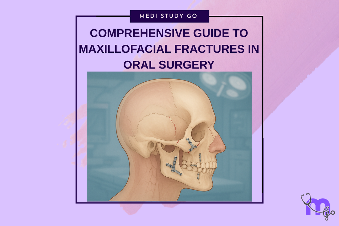
Comprehensive Guide to Maxillofacial Fractures in Oral Surgery
Medi Study Go
Related Resources
Le Fort Fractures: Classification, Clinical Features, and Management
Classification of Midfacial Fractures: Systems and Exam Tips
Zygomatic Complex Fractures: Diagnosis and Surgical Approaches
Gillies Temporal Approach in Zygomatic Arch Fractures
Orbital Blowout Fractures: Pathophysiology and Treatment Protocols
Mandibular Fractures: Classification Systems and Clinical Relevance
Champy's Lines of Osteosynthesis: Principles and Application
Mandibular Angle Fractures: Diagnosis, Complications, and Surgical Management
Condylar Fractures of the Mandible: Types, Indications for Surgery, and Outcomes
Basic Principles of Fracture Fixation in Maxillofacial Surgery
Dental Wiring Techniques in Maxillofacial Fracture Management
CSF Rhinorrhea in Maxillofacial Trauma: Causes, Diagnosis, and Management
Epistaxis Associated with Facial Fractures: Emergency Management
Complications of Maxillofacial Fractures: Early and Late Sequelae
Key Takeaways
- Maxillofacial fractures require immediate stabilization following the ABCDE approach, with airway management as the primary concern
- Understanding anatomical force lines and buttress systems is crucial for proper fracture reduction and fixation
- Modern treatment emphasizes rigid internal fixation over prolonged intermaxillary fixation to promote early functional recovery
- Complications can be minimized through proper surgical technique, adequate reduction, and appropriate fixation methods
- Timing of intervention significantly impacts functional and aesthetic outcomes
Introduction
Maxillofacial fractures represent one of the most challenging aspects of oral and maxillofacial surgery, requiring comprehensive understanding of complex anatomical relationships, biomechanical principles, and advanced surgical techniques. These injuries frequently result from motor vehicle accidents, interpersonal violence, sports injuries, and falls, with significant implications for both function and aesthetics.
The management of maxillofacial fractures has evolved dramatically over the past several decades. While historical approaches relied heavily on prolonged intermaxillary fixation and external immobilization, contemporary treatment emphasizes anatomic reduction, stable internal fixation, and early return to function. This paradigm shift has been driven by improved understanding of fracture healing biology, advances in biomaterials, and recognition of the importance of preserving facial aesthetics and function.
The facial skeleton serves multiple critical functions beyond structural support. It houses and protects vital sensory organs, provides the framework for facial expression, enables mastication and speech, and largely determines facial appearance. Consequently, maxillofacial fractures can have profound effects on a patient's quality of life, affecting not only physical function but also psychological well-being and social interaction.
Modern maxillofacial trauma management requires a multidisciplinary approach, often involving oral and maxillofacial surgeons, neurosurgeons, ophthalmologists, otolaryngologists, and plastic surgeons. The complexity of these injuries demands thorough understanding of anatomical relationships, fracture patterns, healing principles, and reconstruction techniques.
This comprehensive guide examines the fundamental principles of maxillofacial fracture management, from initial assessment and diagnosis through definitive treatment and long-term follow-up. We explore classification systems, diagnostic protocols, treatment planning considerations, and surgical techniques that form the foundation of contemporary practice in maxillofacial trauma surgery.
Table of Contents
- Anatomical Foundations and Biomechanical Principles
- Classification Systems and Fracture Patterns
- Initial Assessment and Emergency Management
- Diagnostic Imaging and Clinical Evaluation
- Treatment Planning and Surgical Approaches
- Complications and Management Strategies
Anatomical Foundations and Biomechanical Principles
The facial skeleton consists of paired and unpaired bones that form a complex three-dimensional framework supporting vital structures and determining facial morphology. Understanding the anatomical relationships and biomechanical properties of these structures is fundamental to successful fracture management.
Structural Organization and Force Distribution
The midface is organized around vertical and horizontal buttresses that distribute masticatory and impact forces throughout the facial skeleton. The vertical buttresses include the nasomaxillary, zygomaticomaxillary, and pterygomaxillary pillars, while horizontal buttresses comprise the frontal bar, infraorbital rims, maxillary alveolar process, and mandibular body.
These buttress systems function as load-bearing columns that transfer forces from the teeth and temporomandibular joints to the skull base. During mastication, forces generated can exceed 200 pounds per square inch in the molar region, requiring robust structural support. The buttress arrangement also provides resistance to impact forces, though severe trauma can overwhelm these protective mechanisms.
Mandibular Biomechanics and Muscle Forces
The mandible functions as a class III lever system, with the temporomandibular joint serving as the fulcrum, muscles of mastication providing the effort, and occlusal contacts representing the load. This arrangement generates significant mechanical advantage for crushing and grinding food but also creates complex force patterns during fracture healing.
Muscle forces acting on fractured mandibular segments can cause significant displacement if not properly controlled. The masseter, temporalis, and medial pterygoid muscles tend to elevate and medialize fracture segments, while the lateral pterygoid muscle causes anterior displacement. The suprahyoid muscles exert downward and backward forces on the mandibular body, particularly in symphyseal and parasymphyseal fractures.
Healing Biology and Fixation Principles
Maxillofacial bone healing follows predictable biological processes involving inflammatory, reparative, and remodeling phases. The rich vascular supply of facial bones generally promotes rapid healing, with most fractures achieving clinical union within 4-6 weeks. However, factors such as infection, poor reduction, inadequate fixation, and patient compliance can significantly affect healing outcomes.
Modern fixation principles emphasize anatomic reduction, stable internal fixation, and preservation of periosteal blood supply. Rigid internal fixation allows immediate function while maintaining fracture stability, promoting better bone healing and reducing complications associated with prolonged immobilization.
What are the main types of maxillofacial fracture classification systems?
Classification systems for maxillofacial fractures serve multiple purposes: they facilitate communication between clinicians, guide treatment planning, predict complications, and enable comparison of treatment outcomes. The most widely used systems include anatomical classifications that describe fracture location and morphological classifications that characterize fracture patterns.
For midface fractures, the Le Fort classification remains the most recognized system, describing three distinct fracture patterns based on anatomical weakness areas. Le Fort I fractures separate the dentoalveolar segment from the rest of the maxilla, Le Fort II fractures create a pyramidal segment including the nasal bones and medial orbital walls, and Le Fort III fractures completely separate the midface from the cranial base.
However, pure Le Fort fractures are relatively uncommon in clinical practice. Most midface fractures involve combinations of patterns or unilateral presentations. Contemporary classification systems increasingly emphasize functional implications and treatment requirements rather than purely anatomical descriptions.
Mandibular fracture classification typically focuses on anatomical location, including symphyseal, parasymphyseal, body, angle, ramus, coronoid, and condylar regions. Additional descriptors address fracture favorability, displacement, comminution, and associated injuries. The concept of favorable versus unfavorable fractures considers muscle force vectors and their tendency to maintain reduction or cause displacement.
Classification Systems and Fracture Patterns
Midface Fracture Classifications
The complex anatomy of the midface has led to development of multiple classification systems, each emphasizing different aspects of fracture morphology and clinical significance. The traditional Le Fort classification, described by René Le Fort in 1901 based on cadaveric studies, identifies three horizontal fracture patterns corresponding to lines of structural weakness in the facial skeleton.
Marciani's modification of the Le Fort classification adds subtypes that recognize the frequent occurrence of associated nasal and naso-orbito-ethmoid fractures. These modifications better reflect the reality of clinical practice, where pure Le Fort patterns are uncommon and combination injuries are the norm.
Rowe and Williams proposed a functional classification distinguishing fractures that involve dental occlusion from those that do not. This system recognizes the critical importance of occlusal relationships in treatment planning and emphasizes the different approaches required for fractures affecting masticatory function.
Zygomatic Complex Fractures
Zygomatic complex fractures, often called tripod or tetrapod fractures, involve disruption of multiple suture lines connecting the zygoma to adjacent facial bones. The Knight and North classification describes six groups based on fracture displacement and rotation patterns, providing guidance for treatment planning and surgical approach selection.
More recently, the Rowe and Killey classification focuses on the rotational component of zygomatic fractures, recognizing that most displacement occurs around predictable axes. This understanding has improved surgical techniques for fracture reduction and fixation.
Orbital Fractures
Orbital fractures require specialized classification systems due to their complex three-dimensional anatomy and potential for vision-threatening complications. The distinction between pure blowout fractures (isolated orbital wall fractures with intact orbital rim) and impure fractures (combined wall and rim involvement) has important implications for surgical approach and timing.
Manson's classification of orbital fractures considers the energy of impact and degree of comminution, helping predict surgical complexity and potential complications. This system recognizes that high-energy injuries often involve multiple orbital walls and require more extensive reconstruction.
Initial Assessment and Emergency Management
Primary Survey and Life-Threatening Conditions
The initial management of maxillofacial trauma follows established trauma protocols, beginning with the primary survey addressing airway, breathing, circulation, disability, and exposure. Maxillofacial injuries can compromise the airway through multiple mechanisms, including bleeding, tissue swelling, bony displacement, and loss of anatomical landmarks.
Airway obstruction may be immediately life-threatening and requires prompt recognition and intervention. Signs of airway compromise include stridor, altered voice, difficulty swallowing, and respiratory distress. Bleeding from facial fractures can be profuse, particularly with Le Fort fractures and complex midface injuries, requiring aggressive fluid resuscitation and hemorrhage control.
How should airway management be approached in maxillofacial trauma?
Airway management in maxillofacial trauma requires careful assessment of injury patterns and available intervention options. Conscious patients with intact neurological function often maintain their own airway most effectively through positioning and natural protective reflexes. However, deterioration can occur rapidly due to progressive swelling or continued bleeding.
When airway intervention is required, options include nasotracheal intubation, orotracheal intubation with direct or video laryngoscopy, supraglottic airway devices, and surgical airway establishment. The choice depends on injury severity, anatomical distortion, operator experience, and available equipment.
Nasotracheal intubation should be avoided in patients with suspected skull base fractures, nasal fractures with significant displacement, or evidence of cerebrospinal fluid rhinorrhea. Orotracheal intubation may be challenging due to trismus, dental injuries, or mandibular fractures but remains the preferred approach in most situations.
Diagnostic Imaging and Clinical Evaluation
Clinical Assessment Techniques
Systematic clinical evaluation begins with inspection for obvious deformities, asymmetries, and soft tissue injuries. Palpation should assess for step defects, crepitus, and abnormal mobility while being gentle enough to avoid further injury. Functional assessment includes evaluation of occlusion, mandibular range of motion, and cranial nerve function.
Specific clinical tests can help identify particular fracture patterns. The McGrigor-Campbell lines provide a systematic approach to assessing midface fractures on Waters radiographs, while manual palpation of suspected fracture sites can reveal characteristic findings such as step defects and crepitus.
Advanced Imaging Protocols
Computed tomography has revolutionized the diagnosis and treatment planning for maxillofacial fractures. Multiplanar imaging with fine-cut sections allows precise visualization of fracture patterns, displacement, and associated injuries. Three-dimensional reconstructions facilitate surgical planning and patient education.
Modern CT protocols typically include axial, coronal, and sagittal reformations with soft tissue and bone window settings. Specific attention should be paid to orbital contents in periorbital fractures, assessing for muscle entrapment or globe injury. Vascular imaging may be indicated for high-energy injuries with potential vessel involvement.
What imaging studies are most appropriate for different fracture types?
The selection of appropriate imaging studies depends on clinical findings, suspected injury patterns, and treatment planning requirements. Simple mandibular fractures may be adequately assessed with panoramic radiography and selected plain films, while complex midface injuries typically require CT evaluation.
Magnetic resonance imaging is occasionally useful for assessing soft tissue injuries, particularly orbital contents, extraocular muscles, and optic nerve pathology. However, CT remains the gold standard for bone injury evaluation and surgical planning in most cases.
For condylar fractures, MRI may provide additional information about disc position and joint space relationships, particularly when considering surgical intervention. However, the clinical utility of this information in treatment decision-making remains debated.
Treatment Planning and Surgical Approaches
Principles of Fracture Reduction and Fixation
Modern maxillofacial fracture treatment is based on principles of anatomic reduction, stable fixation, and early return of function. The goals include restoration of facial height, width, and projection, reestablishment of dental occlusion, and preservation of sensory and motor nerve function.
The sequence of fracture repair typically follows the principle of restoring facial height first, then width, and finally projection. This approach helps ensure proper three-dimensional relationships and facilitates accurate reduction of subsequent fractures. In complex panfacial injuries, establishing stable reference points early in the reconstruction guides subsequent repair.
Fixation Methods and Materials
Contemporary fixation methods include transosseous wiring, miniplates and screws, and reconstruction plates. The choice depends on fracture location, pattern, patient factors, and surgeon preference. Miniplate fixation has become the standard for most facial fractures due to its versatility, strength, and biocompatibility.
Plate positioning follows established principles such as Champy's lines for mandibular fractures, which identify optimal locations for load-sharing fixation. These positions correspond to areas of maximum tensile stress and provide effective resistance to deforming forces while minimizing hardware bulk and prominence.
Surgical Approaches and Access
Surgical access to facial fractures requires careful consideration of cosmetic outcomes, functional preservation, and adequate exposure. Approaches may be intraoral, extraoral, or combined, depending on fracture location and complexity.
Intraoral approaches offer excellent cosmesis but may provide limited access for complex reductions or fixation. Common intraoral incisions include vestibular incisions for mandibular and maxillary fractures and sublingual approaches for lingual plate application.
Extraoral approaches provide superior visualization and access but create visible scars and risk injury to facial nerve branches. Popular extraoral approaches include coronal incisions for upper facial access, preauricular approaches for zygomatic arch fractures, and submandibular incisions for mandibular angle and body fractures.
Complications and Management Strategies
Early Complications
Early complications of maxillofacial fractures include infection, hemorrhage, nerve injury, and dental complications. Infection risk is elevated in open fractures, particularly those involving the oral cavity, and requires aggressive antibiotic therapy and surgical debridement when indicated.
Hemorrhage may be immediate or delayed and can originate from injured vessels, soft tissues, or bone surfaces. Massive bleeding from midface fractures may require emergency arterial ligation or embolization. The sphenopalatine and maxillary arteries are common sources of significant bleeding in Le Fort fractures.
Nerve injuries are common in facial fractures, with the infraorbital, mental, and marginal mandibular nerves being particularly vulnerable. While most nerve injuries are temporary due to neuropraxia, some result in permanent dysfunction requiring additional treatment or acceptance of functional deficits.
Late Complications and Secondary Reconstruction
Late complications include malunion, nonunion, chronic pain, and functional deficits. Malunion is often the result of inadequate reduction, loss of fixation, or premature mobilization. Prevention through proper surgical technique and patient compliance is preferable to secondary correction.
Nonunion is uncommon in facial fractures due to the excellent blood supply but may occur in infected fractures, pathological fractures, or cases with severe soft tissue loss. Treatment typically requires debridement of nonviable tissue, bone grafting, and rigid internal fixation.
How can complications be prevented in maxillofacial fracture treatment?
Prevention of complications begins with proper initial assessment, appropriate treatment planning, and meticulous surgical technique. Key preventive measures include achieving adequate reduction, selecting appropriate fixation methods, preserving soft tissue attachments, and maintaining sterile technique.
Patient education and compliance are crucial for preventing complications. Patients must understand activity restrictions, oral hygiene requirements, and signs of potential problems requiring immediate attention. Regular follow-up allows early detection and treatment of developing complications.
Antibiotic prophylaxis is generally recommended for open fractures or those requiring intraoral approaches. The duration and selection of antibiotics should follow established protocols while considering individual patient factors and local resistance patterns.
Conclusion
Maxillofacial fracture management represents one of the most challenging and rewarding aspects of oral and maxillofacial surgery. Success requires comprehensive understanding of anatomical relationships, biomechanical principles, and healing biology, combined with technical surgical skills and clinical judgment.
The evolution from prolonged immobilization to rigid internal fixation has dramatically improved outcomes for patients with facial fractures. Contemporary treatment emphasizes anatomic restoration, functional preservation, and aesthetic optimization through careful surgical planning and execution.
Future developments in maxillofacial trauma care will likely focus on improved biomaterials, computer-assisted surgery, and regenerative techniques. However, the fundamental principles of anatomic reduction, stable fixation, and early function will remain central to successful treatment.
The complexity of maxillofacial fractures demands ongoing education and skill development. Surgeons must stay current with evolving techniques, technologies, and best practices while maintaining focus on patient-centered care and optimal outcomes. Through continued advancement in understanding and technique, we can further improve the lives of patients affected by facial trauma.