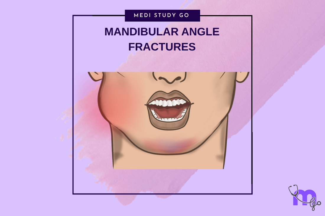
Mandibular Angle Fractures: Diagnosis, Complications, and Surgical Management
Medi Study Go
Related Resources
Comprehensive Guide to Maxillofacial Fractures in Oral Surgery
Le Fort Fractures: Classification, Clinical Features, and Management
Classification of Midfacial Fractures: Systems and Exam Tips
Zygomatic Complex Fractures: Diagnosis and Surgical Approaches
Gillies Temporal Approach in Zygomatic Arch Fractures
Orbital Blowout Fractures: Pathophysiology and Treatment Protocols
Mandibular Fractures: Classification Systems and Clinical Relevance
Champy's Lines of Osteosynthesis: Principles and Application
Condylar Fractures of the Mandible: Types, Indications for Surgery, and Outcomes
Basic Principles of Fracture Fixation in Maxillofacial Surgery
Dental Wiring Techniques in Maxillofacial Fracture Management
CSF Rhinorrhea in Maxillofacial Trauma: Causes, Diagnosis, and Management
Epistaxis Associated with Facial Fractures: Emergency Management
Complications of Maxillofacial Fractures: Early and Late Sequelae
Key Takeaways
- Mandibular angle fractures are the most common mandibular fractures due to anatomical weakness
- Third molar presence significantly increases fracture risk and influences treatment complexity
- Favorable versus unfavorable fracture patterns determine treatment approach and prognosis
- Single miniplate fixation along Champy's line provides adequate stability for most cases
- Complication rates are higher than other mandibular fracture locations
Introduction
Mandibular angle fractures represent the most frequently encountered mandibular fractures, accounting for approximately 30-40% of all mandibular injuries. The unique anatomical characteristics of the mandibular angle region create a relative weakness that predisposes to fracture while simultaneously presenting complex treatment challenges due to difficult surgical access and high complication rates.
The mandibular angle extends from the anterior border of the masseter muscle to the posterior border of the mandible, encompassing the region where the horizontal body transitions to the vertical ramus. This anatomical transition zone, combined with the frequent presence of impacted third molars, creates biomechanical and structural vulnerabilities that explain the high fracture incidence.
Understanding mandibular angle fractures requires comprehensive knowledge of regional anatomy, biomechanical principles, and the complex interplay between fracture pattern, third molar status, and treatment selection. The management of these fractures has evolved significantly with the development of rigid internal fixation techniques and improved understanding of complications.
Anatomical Considerations and Etiology
Regional Anatomy
The mandibular angle region represents a transition zone between the horizontal body and vertical ramus of the mandible. This area has reduced cross-sectional area compared to adjacent regions, creating a relative weakness that predisposes to fracture under stress.
The presence of the mandibular third molar further weakens this region by reducing available bone cross-sectional area. Impacted third molars create additional stress concentration points and may alter the fracture pattern when injuries occur.
Biomechanical Factors
The angle region experiences complex force patterns during mandibular function. The masseter muscle attachment creates compressive forces on the lateral aspect, while the medial pterygoid muscle produces similar forces medially. These opposing forces can cause fracture displacement if not properly controlled during treatment.
The curved anatomy of the angle region also creates rotational forces that tend to displace fracture segments. Understanding these biomechanical factors is crucial for successful treatment planning and fracture reduction.
Classification and Fracture Patterns
Anatomical Classification
Mandibular angle fractures are classified based on their relationship to the third molar and the direction of fracture line propagation. Fractures may occur anterior to, through, or posterior to the third molar socket, with each location presenting different treatment considerations.
The relationship to the third molar influences fracture stability, treatment complexity, and complication risk. Fractures through the third molar socket often require tooth extraction and may have higher infection rates.
What determines favorable versus unfavorable angle fractures?
Favorable angle fractures have fracture lines that resist displacement by muscle forces, typically running in a vertical or near-vertical direction. The fracture geometry allows muscle forces to compress rather than separate the fracture segments.
Unfavorable fractures have oblique or horizontal fracture lines that promote displacement despite reduction efforts. These fractures tend to gap under muscle forces and require more robust fixation to maintain reduction.
Horizontal vs. Vertical Patterns
Horizontal fracture lines are generally unfavorable because they tend to separate under the influence of elevator muscles. Vertical fracture lines may be favorable if properly reduced, as muscle forces tend to compress the fracture site during function.
The angle of fracture line inclination significantly affects treatment difficulty and prognosis. Severely unfavorable patterns may require multiple fixation points or alternative treatment approaches.

Clinical Presentation and Diagnosis
Signs and Symptoms
Patients with mandibular angle fractures present with characteristic signs and symptoms including pain, swelling, trismus, malocclusion, and altered sensation in the distribution of the inferior alveolar nerve. The severity of symptoms often correlates with fracture displacement and associated injuries.
Trismus may be severe due to masseter muscle spasm and mechanical interference from displaced fracture segments. The limitation of mandibular opening can complicate clinical examination and anesthesia administration.
Physical Examination
Clinical examination reveals step defects along the inferior border of the mandible, tenderness over the angle region, and possible crepitus on palpation. Intraoral examination may reveal mucosal lacerations, dental injuries, and occlusal disturbances.
The Coleman sign (sublingual ecchymosis) is often present in mandibular fractures and represents bleeding from the fracture site into the floor of the mouth. This finding, while not specific to angle fractures, supports the diagnosis of mandibular injury.
Imaging Studies
Panoramic radiography provides excellent visualization of mandibular angle fractures and is often the initial imaging study of choice. CT scanning offers superior detail for complex fractures and surgical planning, particularly for comminuted or displaced fractures.
Three-dimensional reconstructions help visualize fracture patterns and displacement, facilitating surgical planning and patient education. The relationship between fracture lines and the third molar is best assessed with high-resolution CT imaging.
Treatment Principles and Decision Making
Conservative vs. Surgical Management
The decision between conservative and surgical management depends on fracture displacement, occlusal disturbance, and patient factors. Minimally displaced favorable fractures in dentate patients may be managed with intermaxillary fixation alone.
Most angle fractures require surgical intervention due to displacement, unfavorable fracture patterns, or functional deficits. The trend toward rigid internal fixation has improved outcomes and reduced treatment times compared to conservative management.
Surgical Approaches
The choice of surgical approach depends on fracture pattern, surgeon preference, and associated injuries. Intraoral approaches through the external oblique ridge provide good access while avoiding external scars but may have higher infection rates.
Extraoral approaches through submandibular or Risdon incisions offer excellent visualization and lower infection rates but create external scars and risk marginal mandibular nerve injury.
How should third molars be managed in angle fractures?
Third molars in the fracture line should generally be removed if they interfere with reduction, show signs of pathology, or prevent adequate fixation placement. Healthy third molars not in the fracture line may be preserved.
The decision requires careful evaluation of tooth position, periodontal status, and fracture pattern. Removal of healthy teeth outside the fracture line is generally not indicated unless they complicate treatment.
Surgical Techniques and Fixation Methods
Miniplate Fixation
Single miniplate fixation along Champy's line (external oblique ridge) provides adequate stability for most mandibular angle fractures. The plate should be positioned as high as possible while avoiding third molar roots and maintaining adequate bone purchase.
Two-point fixation may be necessary for severely unfavorable fractures or those with significant comminution. The second plate is typically placed along the inferior border, maintaining adequate separation from the superior plate.
Screw Selection and Placement
Monocortical screws are typically adequate for angle fracture fixation when plates are properly positioned. Bicortical screws may be necessary in cases with poor bone quality or unstable fracture patterns.
Screw placement requires careful attention to anatomical structures, particularly the inferior alveolar nerve and adjacent tooth roots. Proper drilling technique and screw selection minimize the risk of complications.
Alternative Fixation Methods
Traditional methods including closed reduction with intermaxillary fixation remain viable options for select cases. Transosseous wiring techniques may be useful in cases where plate fixation is not feasible.
External fixation devices are rarely used for angle fractures but may be considered in cases with severe contamination or when internal fixation is contraindicated.
Complications and Management
Early Complications
Mandibular angle fractures have higher complication rates than other mandibular fracture locations. Common early complications include infection, dehiscence, nerve injury, and malocclusion.
Infection rates are particularly high due to bacterial contamination from the oral cavity and the presence of third molars in the fracture site. Aggressive antibiotic therapy and surgical debridement may be necessary.
Late Complications
Late complications include malunion, nonunion, chronic pain, and temporomandibular joint dysfunction. These complications may require extensive secondary reconstruction procedures.
Hardware-related complications such as plate exposure, loosening, or fracture may occur months to years after initial treatment. Plate removal may be necessary in symptomatic cases after adequate healing.
Prevention Strategies
Complication prevention begins with appropriate case selection, proper surgical technique, and postoperative care. Factors contributing to complications include smoking, poor oral hygiene, medical comorbidities, and technical errors.
Patient education and compliance with postoperative instructions significantly influence complication rates. Regular follow-up allows early detection and management of developing problems.
Special Considerations
Pediatric Patients
Mandibular angle fractures in children present unique challenges due to developing dentition and growth considerations. Conservative management is often preferred when possible to avoid growth disturbances.
When surgical intervention is necessary, careful attention to tooth buds and growth centers is essential. Hardware removal may be indicated after healing to prevent growth interference.
Edentulous Patients
Angle fractures in edentulous patients may be more challenging to treat due to bone atrophy and lack of dental landmarks. Fracture patterns may differ from those in dentate patients due to altered biomechanics.
Treatment may require different approaches and fixation techniques adapted to the altered anatomy. The use of reconstruction plates may be necessary in severely atrophic mandibles.
Outcomes and Prognosis
Functional Results
Most patients achieve satisfactory functional outcomes following appropriate treatment of mandibular angle fractures. Return to normal diet and jaw function typically occurs within 6-8 weeks for uncomplicated cases.
Long-term studies show good maintenance of dental occlusion and jaw function in successfully treated cases. However, some patients may experience persistent symptoms such as mild trismus or altered sensation.
Factors Affecting Outcomes
Several factors influence treatment outcomes including patient age, fracture pattern, treatment method, and compliance with postoperative care. Favorable fractures generally have better outcomes than unfavorable patterns.
The presence of complications significantly affects outcomes and may require additional treatment or result in permanent functional deficits. Prevention through proper technique and patient selection is crucial.
Conclusion
Mandibular angle fractures represent challenging injuries that require comprehensive understanding of anatomy, biomechanics, and treatment principles. The high complication rates associated with these fractures emphasize the importance of careful treatment planning and meticulous surgical technique.
Modern treatment approaches utilizing rigid internal fixation have improved outcomes compared to historical methods. However, the fundamental principles of fracture reduction, stable fixation, and prevention of complications remain central to successful management.
Future developments in treatment techniques, materials, and understanding of healing biology will likely further improve outcomes for patients with mandibular angle fractures. The emphasis on evidence-based treatment and complication prevention will continue to guide clinical practice evolution.