Orbital Blowout Fractures: Pathophysiology and Treatment Protocols
Medi Study Go
Related Resources
Comprehensive Guide to Maxillofacial Fractures in Oral Surgery
Le Fort Fractures: Classification, Clinical Features, and Management
Classification of Midfacial Fractures: Systems and Exam Tips
Zygomatic Complex Fractures: Diagnosis and Surgical Approaches
Gillies Temporal Approach in Zygomatic Arch Fractures
Mandibular Fractures: Classification Systems and Clinical Relevance
Champy's Lines of Osteosynthesis: Principles and Application
Mandibular Angle Fractures: Diagnosis, Complications, and Surgical Management
Condylar Fractures of the Mandible: Types, Indications for Surgery, and Outcomes
Basic Principles of Fracture Fixation in Maxillofacial Surgery
Dental Wiring Techniques in Maxillofacial Fracture Management
CSF Rhinorrhea in Maxillofacial Trauma: Causes, Diagnosis, and Management
Epistaxis Associated with Facial Fractures: Emergency Management
Complications of Maxillofacial Fractures: Early and Late Sequelae
Key Takeaways
- Orbital blowout fractures result from hydraulic pressure mechanisms causing orbital wall failure
- Early ophthalmologic evaluation is critical for detecting vision-threatening complications
- Surgical intervention is indicated for persistent diplopia, enophthalmos, or muscle entrapment
- CT imaging with fine-cut sections provides optimal visualization for surgical planning
- Timely treatment within 2 weeks optimizes functional outcomes and prevents complications
Introduction
Orbital blowout fractures represent a unique subset of facial trauma characterized by fractures of the orbital walls with intact orbital rims. These injuries result from specific mechanisms that create isolated orbital wall failures, leading to characteristic clinical presentations and treatment challenges. Understanding the pathophysiology, diagnostic criteria, and treatment protocols is essential for optimal patient management.
The term "blowout" was coined to describe the mechanism by which increased intraorbital pressure causes fracture of the weakest orbital walls, typically the floor and medial wall. This concept explains the characteristic patterns seen in these injuries and guides both diagnostic and therapeutic approaches.
Modern management of orbital blowout fractures requires multidisciplinary collaboration between maxillofacial surgeons, ophthalmologists, and radiologists. The complex anatomy of the orbit and its contents demands comprehensive evaluation and careful treatment planning to achieve optimal functional and aesthetic outcomes.
Pathophysiology and Mechanisms

Hydraulic Theory
The hydraulic theory explains orbital blowout fractures as the result of sudden increases in intraorbital pressure following blunt trauma. When a large object strikes the orbit, the pressure is transmitted through the orbital contents, causing fracture at the weakest points of the orbital walls.
The orbital floor, particularly in the posteromedial region, represents the thinnest and weakest area of the orbital walls. This anatomical vulnerability explains the frequent involvement of the orbital floor in blowout fractures.
Buckling Mechanism
An alternative explanation involves the buckling mechanism, where force transmission through the orbital rim causes deformation and fracture of the orbital walls. This mechanism may explain fractures that occur with rim involvement or in cases where direct pressure transmission seems unlikely.
Both mechanisms likely contribute to different injury patterns, and understanding these concepts helps predict fracture locations and associated injuries.
What anatomical factors predispose to orbital blowout fractures?
The anatomy of the orbital floor creates a natural predisposition to blowout fractures. The posteromedial floor, overlying the maxillary sinus, is particularly thin and lacks substantial bony support. This area, often called the "blowout zone," represents the most common site of orbital floor fractures.
The infraorbital groove and canal create additional areas of weakness where fractures commonly propagate. The relationship between orbital contents and these anatomical variations influences both fracture patterns and clinical presentations.
Classification and Types
Pure vs. Impure Blowout Fractures
Pure blowout fractures involve isolated orbital wall fractures with intact orbital rims. These injuries typically result from small object impacts that create pressure increases without rim fractures. Examples include sports balls, fists, or other rounded objects.
Impure blowout fractures involve orbital wall fractures with associated orbital rim fractures. These injuries usually result from higher energy impacts and often occur as part of more complex facial fracture patterns.
Location-Based Classification
Orbital blowout fractures are classified by location: floor, medial wall, roof, or lateral wall fractures. Floor fractures are most common, followed by medial wall fractures. Combined floor and medial wall fractures occur frequently due to the proximity of these structures.
The location influences both clinical presentation and treatment approach. Floor fractures typically cause enophthalmos and vertical diplopia, while medial wall fractures may affect horizontal eye movements.
Clinical Presentation and Diagnosis
Signs and Symptoms
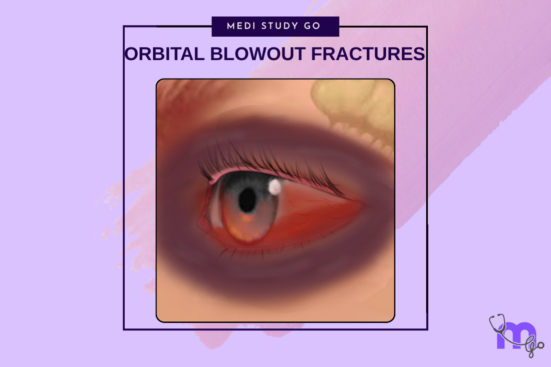
Patients with orbital blowout fractures present with characteristic signs and symptoms that vary depending on fracture location and severity. Common findings include periorbital edema, ecchymosis, subconjunctival hemorrhage, and altered sensation in the infraorbital nerve distribution.
Diplopia represents one of the most significant functional complaints and may result from extraocular muscle entrapment, orbital volume changes, or muscle contusion. The pattern of diplopia helps localize the anatomical structures involved.
Ophthalmologic Evaluation
Comprehensive ophthalmologic evaluation is essential for all suspected orbital fractures. This assessment includes visual acuity testing, pupillary examination, extraocular movement evaluation, and assessment for signs of globe injury or increased intraocular pressure.
The forced duction test helps distinguish muscle entrapment from muscle paresis. Restriction on forced duction suggests mechanical entrapment requiring surgical intervention, while normal forced duction with limited voluntary movement suggests neurological injury.
What clinical findings indicate urgent surgical intervention?
Clinical findings that indicate urgent surgical intervention include persistent diplopia with positive forced duction testing, significant enophthalmos (greater than 2mm), large orbital floor defects (greater than 50% of floor area), and signs of muscle entrapment with nausea and bradycardia.
The "white-eyed blowout fracture" in pediatric patients represents a surgical emergency due to the risk of muscle ischemia and permanent extraocular muscle dysfunction. This condition requires immediate surgical exploration and repair.
Imaging and Radiographic Assessment
CT Imaging Protocols
High-resolution CT scanning with multiplanar reconstructions provides optimal visualization of orbital fractures. Fine-cut sections (1-2mm) in axial and coronal planes allow detailed assessment of fracture extent and orbital contents.
Specific findings to evaluate include orbital floor continuity, herniation of orbital contents into the maxillary sinus, extraocular muscle position, and orbital volume measurements. Three-dimensional reconstructions help with surgical planning and patient education.
Advanced Imaging Techniques
MRI may provide additional information about soft tissue injuries, extraocular muscle function, and optic nerve pathology. However, CT remains the gold standard for bone injury assessment and surgical planning.
Dynamic imaging studies, such as CT or MRI during eye movement, may help assess functional deficits and guide treatment decisions in complex cases.
Treatment Decision Making
Conservative Management Indications
Conservative management is appropriate for small orbital floor fractures without significant functional deficits. Patients with minimal diplopia that resolves within the first week, no evidence of muscle entrapment, and enophthalmos less than 2mm may be managed non-surgically.
Close monitoring is essential during conservative treatment, with serial examinations to assess for development of complications or persistent symptoms that might require delayed surgical intervention.
Surgical Indications
Absolute indications for surgical repair include persistent diplopia beyond two weeks with positive forced duction testing, enophthalmos greater than 2mm, large orbital floor defects (>50% of floor area), and evidence of significant muscle entrapment.
Relative indications include patient occupation requiring precise binocular vision, cosmetic concerns about enophthalmos, and patient preference after thorough discussion of risks and benefits.
Surgical Techniques and Approaches
Transconjunctival Approach
The transconjunctival approach provides excellent access to the orbital floor with minimal visible scarring. This approach involves incision through the conjunctiva below the tarsal plate, allowing direct visualization of the orbital floor defect.
Advantages include excellent cosmesis, minimal ectropion risk, and direct access to the posterior orbital floor. The approach requires careful attention to anatomical planes and gentle tissue handling to prevent complications.
Subciliary and Subtarsal Approaches
External approaches through the lower eyelid provide good visualization but carry higher risks of ectropion and visible scarring. The subciliary approach involves incision 2-3mm below the eyelid margin, while the subtarsal approach uses the natural eyelid crease.
These approaches may be preferred for complex fractures requiring extensive reconstruction or when combined with other facial fracture repairs.
How is orbital volume restoration achieved during surgery?
Orbital volume restoration requires accurate assessment of the fracture defect and appropriate implant selection. The goal is to restore normal orbital anatomy while supporting herniated orbital contents and preventing further prolapse.
Common reconstruction materials include titanium mesh, porous polyethylene sheets, and autogenous bone grafts. The choice depends on defect size, surgeon preference, and patient factors. Proper implant positioning and fixation are crucial for successful outcomes.
Complications and Management
Early Complications
Early complications include persistent diplopia, inadequate correction of enophthalmos, implant malposition, and lower eyelid malposition. Diplopia may persist due to incomplete muscle release, inadequate volume restoration, or muscle fibrosis.
Immediate postoperative assessment should evaluate extraocular movements, eyelid position, and signs of complications such as retrobulbar hemorrhage or infection.
Late Complications
Late complications include chronic diplopia, enophthalmos, implant exposure or migration, and eyelid malposition. These complications may require revision surgery and can be challenging to correct completely.
Prevention through meticulous surgical technique and appropriate patient selection is preferable to secondary revision procedures.
Pediatric Considerations
Orbital blowout fractures in children present unique challenges due to differences in anatomy and healing patterns. The "white-eyed blowout fracture" can cause severe complications if not treated emergently.
Children have more elastic bone that may trap extraocular muscles more completely than adult fractures. The oculocardiac reflex is more prominent in pediatric patients and can cause significant bradycardia and nausea.
Outcomes and Prognosis
Functional Outcomes
Most patients achieve satisfactory functional outcomes following appropriate treatment of orbital blowout fractures. Resolution of diplopia occurs in 80-90% of patients when surgery is performed within the optimal time window.
Factors influencing outcomes include fracture size, timing of intervention, surgical technique, and patient compliance with postoperative care. Early intervention generally yields better results than delayed treatment.
Long-term Follow-up
Long-term follow-up is important for detecting late complications and assessing the stability of surgical repair. Most improvement occurs within the first 3-6 months, though continued improvement may occur for up to one year.
Patient education about realistic expectations and potential for residual symptoms is important for satisfaction with treatment outcomes.
Conclusion
Orbital blowout fractures represent complex injuries requiring comprehensive evaluation and careful treatment planning. Understanding the pathophysiology, diagnostic criteria, and treatment options enables optimal patient management and successful outcomes.
The key to successful treatment lies in appropriate patient selection, proper surgical timing, and meticulous surgical technique. Multidisciplinary collaboration between surgeons and ophthalmologists ensures comprehensive care and optimal functional results.
Modern imaging techniques and surgical approaches have significantly improved outcomes for patients with orbital blowout fractures. Continued advances in understanding and technique will further enhance the ability to restore normal orbital function and anatomy.