
Pathological Calcification: Dystrophic vs. Metastatic
Medi Study Go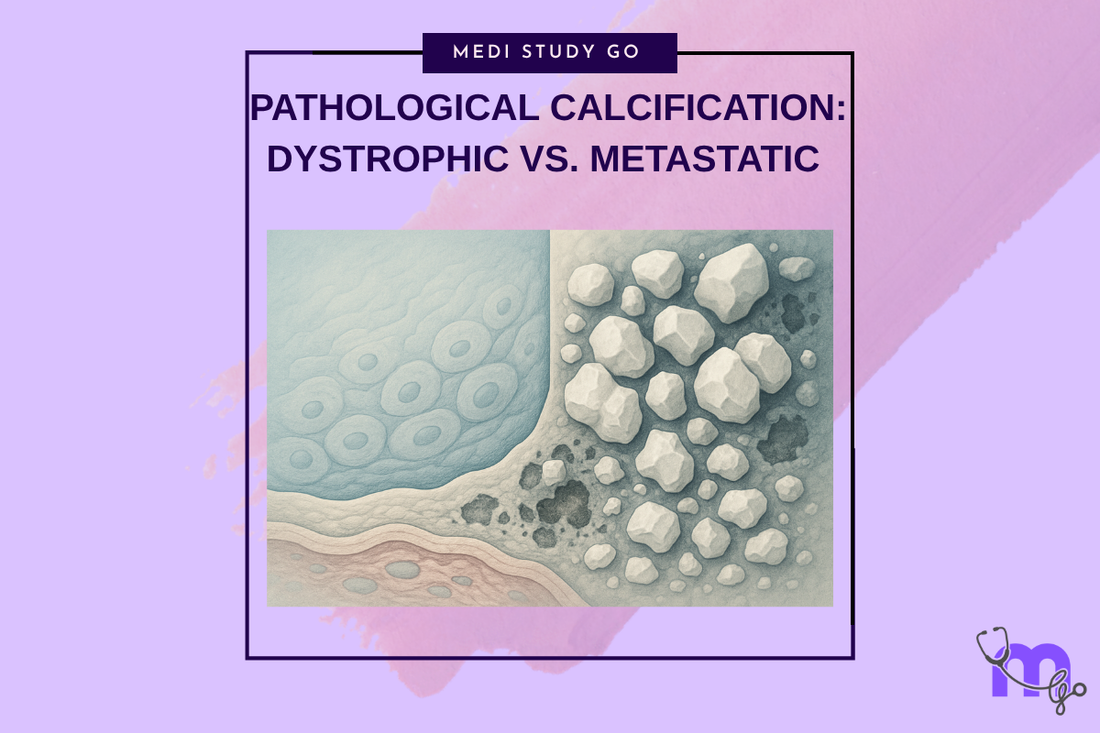
Related Resources
Cell Injury and Adaptation: The Foundation of Pathology
Morphology of Reversible Cell Injury: Key Features and Clinical Examples
Pathogenesis of Reversible Cell Injury: Mechanisms and Outcomes
Pathogenesis of Irreversible Cell Injury: From Damage to Cell Death
Necrosis: Types, Mechanisms, and Clinical Significance
Apoptosis: Programmed Cell Death in Health and Disease
Cellular Adaptation: Types, Mechanisms, and Clinical Relevance
Intracellular Accumulations: Lipids, Proteins, Pigments, and More
Cell Injury in Clinical Practice in Dentistry
Key Takeaways
- Dystrophic calcification occurs in damaged tissues with normal serum calcium levels, while metastatic calcification occurs in normal tissues with elevated serum calcium
- Recognition of calcification patterns helps distinguish between local tissue damage and systemic metabolic disorders affecting calcium homeostasis
- Pulp stones, salivary gland stones, and periodontal calcification represent common examples of pathological calcification in dental practice
- Understanding calcium metabolism disorders enables appropriate medical referral and coordinated management of patients with systemic conditions
- Clinical management strategies differ significantly between dystrophic and metastatic calcification, requiring accurate diagnosis for optimal outcomes
Introduction
Pathological calcification represents the abnormal deposition of calcium salts in tissues where such deposition does not normally occur or occurs in excessive amounts. For dental professionals, understanding pathological calcification is essential due to its frequent occurrence in oral tissues and its potential association with both local pathological processes and systemic metabolic disorders.
The two primary types of pathological calcification demonstrate fundamentally different mechanisms and clinical implications. Dystrophic calcification occurs in previously damaged or necrotic tissues despite normal serum calcium and phosphate levels, while metastatic calcification occurs in normal tissues as a result of elevated serum calcium levels from systemic metabolic disorders.
Recognition of calcification patterns provides important diagnostic information that can distinguish between local tissue pathology and systemic disease, guide appropriate treatment decisions, and determine the need for medical consultation or referral. The clinical significance extends beyond the immediate oral findings to include potential identification of underlying systemic conditions.
In dental practice, pathological calcification is encountered in various forms including pulp stones, salivary gland calculi, periodontal calcification, and soft tissue calcification associated with chronic inflammation or trauma. Understanding the mechanisms and clinical significance of these conditions enables appropriate diagnosis and management while recognizing when systemic evaluation may be indicated.
Contemporary research continues to elucidate the molecular mechanisms underlying pathological calcification, providing insights into prevention strategies and therapeutic approaches. These advances offer opportunities for more effective management of calcification-related conditions and better understanding of the relationship between local and systemic factors in calcium deposition.
Table of Contents
Understanding Calcium Homeostasis and Deposition Mechanisms What Distinguishes Dystrophic from Metastatic Calcification? Clinical Manifestations in Oral and Maxillofacial Tissues How Do Systemic Conditions Influence Pathological Calcification? Diagnostic and Management Strategies
Understanding Calcium Homeostasis and Deposition Mechanisms
Normal Calcium Metabolism
Calcium homeostasis involves complex regulatory mechanisms that maintain serum calcium levels within narrow limits through interactions between parathyroid hormone, vitamin D, calcitonin, and target organs including bone, kidneys, and intestines. Understanding these mechanisms is essential for recognizing conditions that predispose to pathological calcification.
Parathyroid hormone regulation responds to decreased serum calcium by increasing bone resorption, enhancing renal calcium reabsorption, and stimulating vitamin D activation to increase intestinal calcium absorption. These mechanisms normally prevent hypocalcemia but can contribute to hypercalcemia when dysregulated.
Vitamin D metabolism plays crucial roles in calcium absorption and utilization, with deficiency or excess affecting calcium homeostasis and potentially contributing to pathological calcification patterns. Understanding vitamin D status is important for evaluating patients with calcification disorders.
Mechanisms of Pathological Deposition
Calcium phosphate precipitation occurs when local conditions favor crystal formation, including altered pH, increased local calcium or phosphate concentrations, presence of nucleation sites, or absence of crystallization inhibitors. These conditions can develop in both damaged tissues and systemically in hypercalcemic states.
Hydroxyapatite crystal formation represents the most common form of pathological calcification, similar to normal bone and tooth mineralization but occurring in inappropriate locations. The crystal structure and composition provide information about the underlying mechanisms and duration of the calcification process.
Matrix vesicle involvement in pathological calcification parallels normal mineralization processes, with membrane-bound vesicles serving as initial sites for calcium phosphate crystal formation. Understanding this mechanism helps explain why certain tissues are more susceptible to calcification than others.
Tissue Factors Influencing Calcification
Tissue damage creates conditions that favor calcium deposition through several mechanisms including release of intracellular phosphate, altered local pH, exposure of nucleation sites, and decreased activity of calcification inhibitors. These changes explain why dystrophic calcification occurs in damaged tissues.
Inflammatory processes can both promote and inhibit calcification through various mechanisms including altered local blood flow, release of inflammatory mediators that affect calcium metabolism, and changes in tissue pH and oxygenation that influence crystal formation.
Aging processes affect tissue susceptibility to calcification through changes in matrix composition, decreased activity of calcification inhibitors, and accumulated tissue damage that provides nucleation sites for calcium deposition. Understanding age-related changes helps predict calcification risk.
What Distinguishes Dystrophic from Metastatic Calcification?
Understanding the fundamental differences between dystrophic and metastatic calcification is crucial for appropriate diagnosis, treatment planning, and recognition of underlying systemic conditions.
Dystrophic Calcification Characteristics
Dystrophic calcification occurs in previously damaged, degenerated, or necrotic tissues despite normal serum calcium and phosphate levels, representing a local tissue response to injury rather than a systemic metabolic disorder. This type of calcification is common in dental practice and typically indicates prior tissue damage.
The mechanism involves local tissue changes that create favorable conditions for calcium deposition, including altered pH, increased phosphate release from damaged cells, exposure of nucleation sites, and decreased activity of calcification inhibitors. These local changes overcome normal inhibitory mechanisms that prevent calcification.
Clinical presentation of dystrophic calcification typically shows a history of prior tissue injury, trauma, or chronic inflammation in the affected area. The calcification appears months to years after the initial injury and may be discovered incidentally during routine radiographic examination.
Common examples in dental practice include pulp stones following pulpal inflammation, calcification in chronically inflamed periapical tissues, salivary gland stones associated with chronic sialadenitis, and soft tissue calcification following trauma or surgical procedures.
Metastatic Calcification Features
Metastatic calcification occurs in normal, undamaged tissues as a result of elevated serum calcium levels from systemic metabolic disorders, representing a systemic rather than local pathological process. This type requires identification and management of underlying systemic conditions.
The mechanism involves supersaturation of extracellular fluids with calcium and phosphate due to systemic metabolic abnormalities, leading to precipitation in tissues that normally have high metabolic activity or alkaline pH conditions that favor crystal formation.
Clinical presentation typically involves multiple tissue sites affected simultaneously, often including organs with high blood flow or metabolic activity such as kidneys, lungs, gastric mucosa, and blood vessels. In the oral cavity, multiple sites may be affected simultaneously.
Underlying systemic conditions include hyperparathyroidism, malignancy-associated hypercalcemia, vitamin D intoxication, chronic renal failure, and various endocrine disorders that disrupt normal calcium homeostasis. Recognition of these conditions is essential for appropriate patient management.
Diagnostic Differentiation
Laboratory evaluation plays a crucial role in distinguishing between dystrophic and metastatic calcification, with serum calcium, phosphate, parathyroid hormone, and vitamin D levels providing essential diagnostic information. Normal values suggest dystrophic calcification while elevated calcium indicates metastatic calcification.
Radiographic patterns may provide clues to the type of calcification, with dystrophic calcification typically showing localized deposits related to sites of prior injury, while metastatic calcification may show more generalized patterns affecting multiple organ systems.
Clinical history and examination help distinguish between these types by identifying prior tissue injury or trauma that would suggest dystrophic calcification versus systemic symptoms that might indicate metabolic disorders causing metastatic calcification.
Clinical Manifestations in Oral and Maxillofacial Tissues
Pulpal and Periapical Calcification
Pulp stones represent common examples of dystrophic calcification occurring in response to pulpal irritation or inflammation from caries, trauma, or restorative procedures. These calcifications can interfere with endodontic procedures and may indicate the need for modified treatment approaches.
Periapical calcification occurs in chronically inflamed periapical tissues, often appearing as radiopaque masses associated with long-standing periapical lesions. Recognition of this calcification helps predict endodontic treatment challenges and may influence treatment planning decisions.
The clinical significance of pulpal calcification includes potential interference with root canal instrumentation, altered pulpal blood flow, and indication of prior pulpal injury that may affect treatment outcomes. Understanding these implications guides appropriate endodontic management strategies.
Salivary Gland Calcification
Salivary gland stones (sialolithiasis) represent dystrophic calcification occurring in response to ductal obstruction, chronic inflammation, or altered saliva composition. These stones can cause significant symptoms and may require surgical removal for symptom resolution.
The pathogenesis involves chronic inflammation or obstruction leading to tissue damage and favorable conditions for calcium deposition, typically around organic debris or inflammatory cells that serve as nucleation sites for crystal formation.
Clinical presentation includes episodic swelling and pain associated with meals, particularly with sour foods that stimulate salivary flow. Chronic cases may show persistent glandular enlargement and decreased salivary function that affects oral health.
Periodontal and Soft Tissue Calcification
Periodontal calcification can occur in response to chronic inflammation, trauma, or following periodontal surgery, appearing as radiopaque deposits in periodontal ligament space or alveolar bone. Recognition of this calcification helps predict periodontal treatment outcomes.
Soft tissue calcification may develop following trauma, injection site reactions, or chronic inflammatory conditions, appearing as palpable nodules or radiopaque deposits in clinical and radiographic examination. These deposits may require surgical removal if symptomatic.
The relationship between calcification and healing processes affects treatment planning and outcome prediction, with extensive calcification potentially indicating compromised healing capacity or need for modified treatment approaches.
How Do Systemic Conditions Influence Pathological Calcification?
Endocrine Disorders and Calcium Metabolism
Primary hyperparathyroidism causes elevated parathyroid hormone levels leading to increased bone resorption, enhanced renal calcium reabsorption, and stimulated vitamin D production, resulting in hypercalcemia that can cause metastatic calcification in multiple organs including oral tissues.
Vitamin D disorders including intoxication or abnormal metabolism can disrupt calcium homeostasis and contribute to pathological calcification. Understanding vitamin D status is important for evaluating patients with unexplained calcification or multiple calcification sites.
Thyroid disorders can affect calcium metabolism through various mechanisms including altered parathyroid hormone sensitivity and changes in bone turnover rates. Recognition of these relationships helps identify patients who may benefit from endocrine evaluation.
Chronic Kidney Disease Effects
Chronic kidney disease disrupts calcium and phosphate homeostasis through decreased phosphate excretion, altered vitamin D metabolism, and secondary hyperparathyroidism, creating conditions that favor pathological calcification in multiple tissues including oral structures.
Dialysis patients demonstrate particular susceptibility to pathological calcification due to altered calcium and phosphate balance, use of calcium-containing medications, and chronic inflammation that may contribute to dystrophic calcification processes.
The management of calcification in kidney disease patients requires coordination with nephrology care and consideration of systemic factors that influence calcium metabolism. Understanding these relationships guides appropriate dental management strategies.
Malignancy-Associated Calcification
Paraneoplastic hypercalcemia represents a common cause of metastatic calcification in cancer patients, resulting from parathyroid hormone-related protein production by tumors or osteolytic metastases that release calcium from bone. Recognition of this condition may lead to cancer diagnosis.
Chemotherapy and radiation effects can contribute to both dystrophic calcification through tissue damage and metastatic calcification through effects on calcium metabolism. Understanding these mechanisms helps predict and manage treatment-related complications.
The relationship between malignancy and calcification requires careful evaluation to distinguish between treatment effects, paraneoplastic syndromes, and coincidental findings. This assessment influences both dental and oncological management decisions.
Diagnostic and Management Strategies
Clinical Assessment Approaches
History taking should focus on identifying potential causes of tissue damage for dystrophic calcification or systemic symptoms suggesting metabolic disorders for metastatic calcification. Prior trauma, chronic inflammation, and medication history provide important diagnostic clues.
Physical examination should assess for multiple calcification sites that might suggest systemic disease, signs of endocrine disorders, and evidence of chronic kidney disease or other systemic conditions that affect calcium metabolism.
Radiographic evaluation provides essential information about calcification location, extent, and pattern that helps distinguish between dystrophic and metastatic types while guiding treatment planning and monitoring progression.
Laboratory Investigation
Serum calcium and phosphate levels represent the most important initial laboratory tests for evaluating pathological calcification, with normal values suggesting dystrophic calcification and elevated calcium indicating potential metastatic calcification requiring further evaluation.
Parathyroid hormone levels help identify hyperparathyroidism as a cause of hypercalcemia and metastatic calcification, while vitamin D levels assess for deficiency or intoxication that may contribute to calcium metabolism disorders.
Additional testing may include kidney function assessment, thyroid function evaluation, and tumor markers if malignancy-associated hypercalcemia is suspected. The extent of testing depends on clinical findings and initial laboratory results.
Treatment and Management
Local management of dystrophic calcification focuses on addressing underlying tissue damage when possible and removing calcified deposits if they cause symptoms or interfere with function. Surgical removal may be necessary for large or symptomatic deposits.
Systemic management of metastatic calcification requires treatment of underlying metabolic disorders causing hypercalcemia, often requiring medical or endocrine consultation for appropriate diagnosis and management of systemic conditions.
Prevention strategies include minimizing tissue damage that predisposes to dystrophic calcification and recognizing early signs of systemic conditions that may cause metastatic calcification. Patient education regarding risk factors and symptoms enhances early detection and management.
Monitoring and Follow-up
Long-term monitoring protocols should assess for progression of existing calcification, development of new calcification sites, and effectiveness of treatment interventions. Regular clinical and radiographic evaluation helps guide ongoing management decisions.
Coordination with medical colleagues is essential for patients with systemic conditions causing metastatic calcification, ensuring appropriate management of underlying disorders while addressing oral manifestations and complications.
Patient education regarding the nature of their calcification disorder, expected outcomes, and warning signs requiring medical attention enhances compliance with treatment recommendations and facilitates early intervention for complications.
Conclusion
Understanding pathological calcification patterns and their underlying mechanisms provides essential knowledge for distinguishing between local tissue pathology and systemic metabolic disorders while guiding appropriate treatment decisions and medical referrals. The distinction between dystrophic and metastatic calcification has important implications for diagnosis, treatment planning, and patient management.
Recognition of calcification in oral tissues may provide the first indication of underlying systemic conditions requiring medical evaluation and treatment. This highlights the important role of dental professionals in comprehensive patient care and early detection of systemic diseases.
Contemporary understanding of calcification mechanisms continues to evolve, providing insights into prevention strategies and therapeutic approaches that may improve outcomes for patients with pathological calcification. These advances offer opportunities for more effective management and better coordination between dental and medical care.
The integration of local and systemic assessment approaches represents the foundation for optimal management of pathological calcification disorders, emphasizing the importance of comprehensive patient evaluation and multidisciplinary care when systemic conditions are identified or suspected.