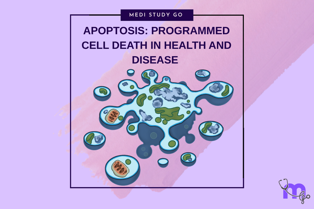
Apoptosis: Programmed Cell Death in Health and Disease
Medi Study Go
Related Resources
Cell Injury and Adaptation: The Foundation of Pathology
Morphology of Reversible Cell Injury: Key Features and Clinical Examples
Pathogenesis of Reversible Cell Injury: Mechanisms and Outcomes
Pathogenesis of Irreversible Cell Injury: From Damage to Cell Death
Necrosis: Types, Mechanisms, and Clinical Significance
Pathological Calcification: Dystrophic vs. Metastatic
Cellular Adaptation: Types, Mechanisms, and Clinical Relevance
Intracellular Accumulations: Lipids, Proteins, Pigments, and More
Cell Injury in Clinical Practice in Dentistry
Key Takeaways
- Apoptosis represents controlled programmed cell death essential for normal development, tissue homeostasis, and elimination of damaged cells
- Recognition of apoptotic vs. necrotic cell death patterns influences treatment decisions and understanding of disease progression in dental conditions
- Apoptosis plays crucial roles in tooth development, periodontal remodeling, and oral tissue turnover throughout life
- Dysregulated apoptosis contributes to various oral pathological conditions including cancer, autoimmune diseases, and healing disorders
- Understanding apoptotic pathways provides insights into therapeutic targets for managing oral diseases and optimizing healing processes
Introduction
Apoptosis represents a highly regulated form of programmed cell death that plays essential roles in normal development, tissue homeostasis, and elimination of damaged or unwanted cells. Unlike necrosis, which represents uncontrolled cell death resulting from severe injury, apoptosis is a controlled process that eliminates cells without causing inflammation or damage to surrounding tissues.
For dental professionals, understanding apoptosis is crucial for comprehending normal oral development, tissue remodeling processes, and various pathological conditions affecting oral tissues. Apoptosis contributes to numerous physiological processes including tooth development and eruption, periodontal ligament remodeling during orthodontic movement, and normal epithelial turnover in oral mucosa.
The molecular mechanisms of apoptosis involve specific signaling pathways that can be triggered by various stimuli including DNA damage, growth factor withdrawal, cellular stress, and developmental signals. These pathways converge on common execution mechanisms that systematically dismantle cellular components while maintaining membrane integrity and preventing inflammatory responses.
Dysregulation of apoptotic processes contributes to various oral pathological conditions, with insufficient apoptosis potentially leading to excessive cell accumulation and neoplastic growth, while excessive apoptosis can result in tissue loss and impaired healing. Understanding these relationships enables better comprehension of disease mechanisms and potential therapeutic approaches.
Contemporary research continues to elucidate the complex regulation of apoptotic pathways and their roles in oral health and disease. These advances provide insights into potential therapeutic targets for managing conditions characterized by dysregulated apoptosis and offer opportunities for enhancing natural healing processes through modulation of programmed cell death pathways.
Table of Contents
Molecular Mechanisms of Apoptotic Cell Death What Distinguishes Apoptosis from Necrosis in Clinical Practice? Physiological Roles of Apoptosis in Oral Development How Does Dysregulated Apoptosis Contribute to Oral Diseases? Therapeutic Implications and Clinical Applications
Molecular Mechanisms of Apoptotic Cell Death
Intrinsic Apoptotic Pathway
The intrinsic apoptotic pathway is triggered by intracellular stress signals including DNA damage, oxidative stress, and growth factor withdrawal, leading to mitochondrial membrane permeabilization and release of pro-apoptotic factors. This pathway is regulated by the Bcl-2 family of proteins, which includes both pro-apoptotic and anti-apoptotic members that determine cellular fate.
Mitochondrial cytochrome c release represents a critical step in the intrinsic pathway, as this protein normally involved in electron transport becomes a signal for apoptosis activation when released into the cytoplasm. The release occurs through mitochondrial outer membrane permeabilization controlled by pro-apoptotic proteins including Bax and Bak.
Apoptosome formation occurs when cytochrome c combines with Apaf-1 and pro-caspase-9 to create a protein complex that activates caspase-9, initiating the caspase cascade that systematically dismantles cellular components. This process represents the point of no return in apoptotic cell death.
Extrinsic Apoptotic Pathway
The extrinsic pathway is triggered by external death signals received through death receptors on the cell surface, including Fas, TNF receptor, and TRAIL receptors. These receptors, when bound by their respective ligands, initiate apoptotic signaling cascades that can rapidly lead to cell death.
Death-inducing signaling complex (DISC) formation occurs when death receptors are activated, leading to recruitment and activation of caspase-8, which can directly activate executioner caspases or amplify the signal through the intrinsic pathway. This mechanism allows for rapid elimination of cells targeted by immune responses or developmental signals.
The regulation of death receptor expression and sensitivity provides important control mechanisms for apoptosis, allowing cells to respond appropriately to external death signals while avoiding inappropriate cell death during normal cellular function.
Execution Phase and Caspase Activation
Caspase cascade activation represents the final common pathway for both intrinsic and extrinsic apoptotic pathways, involving systematic activation of proteolytic enzymes that cleave specific cellular substrates. Executioner caspases including caspase-3, -6, and -7 are responsible for the characteristic morphological and biochemical changes of apoptosis.
Nuclear changes in apoptosis include chromatin condensation, DNA fragmentation into characteristic laddering patterns, and nuclear fragmentation that occurs through caspase-activated DNase activity. These changes distinguish apoptotic from necrotic cell death and provide important diagnostic markers.
Membrane changes during apoptosis include phosphatidylserine externalization, which serves as an "eat-me" signal for phagocytes, and formation of apoptotic bodies that package cellular contents for removal without inflammatory response. These changes enable efficient clearance of apoptotic cells.
What Distinguishes Apoptosis from Necrosis in Clinical Practice?
Understanding the differences between apoptotic and necrotic cell death is crucial for clinical diagnosis, treatment planning, and predicting tissue responses to injury and therapeutic interventions.
Morphological Differences
Apoptotic cells demonstrate characteristic morphological changes including cell shrinkage, chromatin condensation, and formation of apoptotic bodies that maintain membrane integrity throughout the death process. These changes create a distinctive appearance that can be recognized through microscopic examination.
Nuclear morphology in apoptosis shows characteristic pyknosis and fragmentation patterns that differ from the nuclear changes seen in necrosis. Apoptotic nuclei demonstrate organized fragmentation while necrotic nuclei show random degradation and eventual dissolution.
Cellular membrane integrity is maintained in apoptosis until final phagocytic removal, preventing release of cellular contents that could damage surrounding tissues. This contrasts sharply with necrotic cell death where membrane rupture leads to cellular content release and inflammatory responses.
Inflammatory Response Patterns
Apoptotic cell death typically occurs without significant inflammatory response, as the controlled nature of the process and maintenance of membrane integrity prevents release of damage-associated molecular patterns (DAMPs) that trigger inflammation. This characteristic enables tissue remodeling without inflammatory damage.
Phagocytic clearance of apoptotic cells occurs rapidly and efficiently through recognition of phosphatidylserine and other "eat-me" signals, preventing secondary necrosis and inflammatory responses. Professional phagocytes including macrophages and dendritic cells are primarily responsible for this clearance.
The absence of inflammatory response in apoptosis makes this form of cell death ideal for developmental processes, tissue homeostasis, and elimination of potentially harmful cells without tissue damage. Understanding this principle guides therapeutic approaches that promote apoptotic over necrotic cell death.
Clinical Recognition and Implications
Tissue responses to apoptotic cell death include efficient remodeling without scarring or inflammatory damage, making this form of cell death preferable in many clinical situations. Recognition of apoptotic patterns indicates controlled tissue remodeling rather than pathological destruction.
Healing patterns following apoptotic cell death typically show better outcomes with less scarring and more complete tissue restoration compared to healing following necrotic tissue death. This understanding influences treatment approaches that aim to promote apoptotic mechanisms.
The clinical distinction between apoptotic and necrotic patterns helps predict treatment outcomes and guides selection of appropriate therapeutic interventions. Conditions characterized by appropriate apoptosis generally have better prognoses than those involving extensive necrosis.
Physiological Roles of Apoptosis in Oral Development
Tooth Development and Morphogenesis
Apoptosis plays essential roles in tooth development, contributing to proper crown and root morphology through elimination of excess cells and tissue remodeling. The characteristic cuspal patterns and root formation depend on precisely regulated apoptotic processes that sculpt developing tooth structures.
Enamel organ remodeling involves extensive apoptosis of cells that are no longer needed after enamel formation is complete, including stellate reticulum and outer enamel epithelium cells. This process enables proper tooth eruption and establishment of normal crown morphology.
Root development requires apoptotic elimination of Hertwig's epithelial root sheath cells after root dentin formation, allowing proper cementum formation and periodontal ligament attachment. Dysregulation of this process can lead to developmental anomalies and periodontal problems.
Periodontal Development and Remodeling
Periodontal ligament formation and maintenance involve continuous apoptotic remodeling that maintains proper fiber organization and cellular populations. This process enables the periodontal ligament to adapt to changing functional demands throughout life.
Alveolar bone remodeling incorporates apoptotic mechanisms for osteoblast and osteoclast regulation, ensuring appropriate bone formation and resorption in response to functional demands. Understanding these mechanisms is crucial for orthodontic treatment planning and periodontal therapy.
Gingival development and maintenance require apoptotic regulation of epithelial cell turnover, ensuring proper tissue architecture and barrier function while allowing for normal physiological renewal. Disruption of these processes can contribute to periodontal disease development.
Oral Mucosal Homeostasis
Epithelial cell turnover in oral mucosa involves continuous apoptotic elimination of surface cells balanced by basal cell proliferation, maintaining proper tissue thickness and barrier function. This process is essential for oral health and resistance to microbial invasion.
Immune cell regulation in oral tissues requires apoptotic mechanisms for eliminating activated immune cells after inflammatory responses, preventing excessive tissue damage and maintaining immune homeostasis. Dysregulation can contribute to chronic inflammatory conditions.
Salivary gland development and maintenance involve apoptotic processes for ductal formation and acinar cell turnover, ensuring proper glandular architecture and function. Understanding these processes is important for managing salivary gland disorders.
How Does Dysregulated Apoptosis Contribute to Oral Diseases?
Insufficient Apoptosis and Neoplastic Growth
Oral cancer development often involves dysregulation of apoptotic pathways, allowing cells with DNA damage or other abnormalities to survive and proliferate rather than undergo programmed cell death. This contributes to malignant transformation and tumor progression.
p53 mutations, found in many oral cancers, disrupt DNA damage-induced apoptosis, allowing cells with genetic damage to survive and accumulate additional mutations. Understanding this mechanism helps explain cancer development and guides therapeutic approaches.
HPV-related oral cancers demonstrate specific patterns of apoptotic dysregulation through viral protein interference with normal cell death pathways. Recognition of these patterns has implications for diagnosis, treatment, and prevention strategies.
Excessive Apoptosis and Tissue Loss
Periodontal disease progression can involve excessive apoptosis of periodontal ligament cells, contributing to attachment loss and bone destruction. Understanding this mechanism provides insights into disease progression and potential therapeutic targets.
Oral lichen planus and other autoimmune conditions demonstrate excessive keratinocyte apoptosis triggered by immune-mediated mechanisms, leading to tissue damage and clinical symptoms. Recognition of this mechanism guides appropriate treatment approaches.
Medication-related oral complications can involve drug-induced apoptosis of oral epithelial cells or other tissues, contributing to mucositis, ulceration, and healing impairment. Understanding these mechanisms helps predict and manage drug-related oral complications.
Therapeutic Resistance and Treatment Implications
Chemotherapy resistance in oral cancers often involves dysregulation of apoptotic pathways, allowing cancer cells to survive treatments designed to trigger cell death. Understanding these mechanisms guides combination therapy approaches and treatment selection.
Radiation therapy resistance can result from enhanced DNA repair mechanisms or altered apoptotic responses that allow cancer cells to survive radiation-induced damage. Recognition of these mechanisms influences treatment planning and dose selection.
The development of targeted therapies for oral diseases increasingly focuses on modulating apoptotic pathways to restore normal cell death responses in diseased tissues. Understanding these approaches guides selection of appropriate therapeutic options.
Therapeutic Implications and Clinical Applications
Enhancing Apoptosis in Cancer Treatment
Cancer therapy strategies increasingly focus on restoring normal apoptotic responses in malignant cells through various mechanisms including p53 pathway restoration, death receptor activation, and mitochondrial targeting. These approaches offer promise for more effective cancer treatment.
Combination therapies that target multiple apoptotic pathways simultaneously may overcome resistance mechanisms and improve treatment outcomes in oral cancers. Understanding these mechanisms guides rational combination therapy design.
Selective targeting of cancer cells while sparing normal tissues requires understanding of the differences in apoptotic regulation between malignant and healthy cells. This knowledge enables development of more effective and less toxic cancer treatments.
Modulating Apoptosis in Inflammatory Diseases
Anti-inflammatory approaches that promote resolution of inflammation through enhanced apoptosis of inflammatory cells can improve outcomes in chronic oral inflammatory conditions. Understanding these mechanisms guides therapeutic selection.
Corticosteroid therapy effects on apoptosis include both beneficial anti-inflammatory actions and potential adverse effects on tissue healing and immune function. Understanding these mechanisms helps optimize corticosteroid use in oral conditions.
Novel therapeutic approaches targeting specific apoptotic pathways in inflammatory diseases offer promise for more precise treatment with fewer side effects. These developments may improve management of conditions like oral lichen planus and pemphigus.
Tissue Engineering and Regenerative Applications
Tissue engineering strategies must consider apoptotic regulation to ensure proper tissue development and integration while avoiding excessive cell death that could compromise regenerative outcomes. Understanding these mechanisms guides scaffold and growth factor selection.
Stem cell therapy applications require understanding of apoptotic regulation to maintain cell viability during processing and implantation while ensuring appropriate differentiation and integration. These considerations influence cell preparation and delivery protocols.
Gene therapy approaches may target apoptotic pathways to enhance tissue regeneration, promote healing, or eliminate diseased cells. Understanding these mechanisms guides vector design and therapeutic gene selection.
Conclusion
Understanding apoptosis mechanisms and their roles in oral health and disease provides essential knowledge for recognizing normal developmental and homeostatic processes while identifying pathological conditions characterized by dysregulated programmed cell death. The distinction between apoptotic and necrotic cell death patterns has important implications for diagnosis, treatment planning, and outcome prediction.
The physiological roles of apoptosis in oral development and tissue maintenance emphasize the importance of this process for normal oral health, while recognition of dysregulated apoptosis in disease states provides insights into pathogenesis and potential therapeutic targets. Contemporary therapeutic approaches increasingly focus on modulating apoptotic pathways to restore normal tissue function.
Clinical applications of apoptosis research continue to expand, with promising developments in cancer therapy, inflammatory disease management, and regenerative medicine. These advances provide opportunities for more precise and effective treatments that target specific mechanisms of disease while preserving normal tissue function.
Future research in apoptosis mechanisms and therapeutic applications promises to provide even more sophisticated approaches to managing oral diseases and optimizing healing processes. The integration of molecular understanding with clinical practice represents the foundation for advancing oral healthcare toward more personalized and effective therapeutic strategies.