Cell Injury and Adaptation: The Foundation of Pathology
Medi Study Go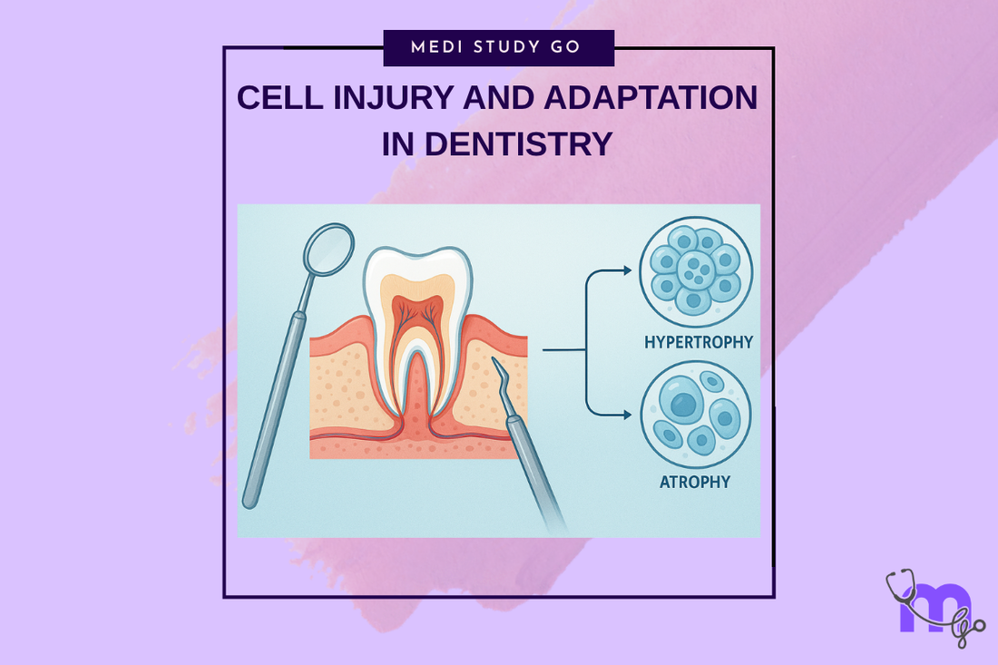
Related Resources
Morphology of Reversible Cell Injury: Key Features and Clinical Examples
Pathogenesis of Reversible Cell Injury: Mechanisms and Outcomes
Pathogenesis of Irreversible Cell Injury: From Damage to Cell Death
Necrosis: Types, Mechanisms, and Clinical Significance
Apoptosis: Programmed Cell Death in Health and Disease
Pathological Calcification: Dystrophic vs. Metastatic
Cellular Adaptation: Types, Mechanisms, and Clinical Relevance
Intracellular Accumulations: Lipids, Proteins, Pigments, and More
Cell Injury in Clinical Practice in Dentistry
Key Takeaways
- Cell injury represents a sequence of events occurring when oral tissues are exposed to stresses exceeding their adaptive capacity, ranging from reversible cellular swelling to irreversible cell death
- Mitochondria, protein synthesis apparatus, cellular membranes, and DNA are the most vulnerable structures to injury in dental tissues, making them critical targets for understanding pathological processes
- Understanding the distinction between reversible and irreversible cell injury is essential for early intervention and optimal treatment outcomes in dental practice
- Cellular adaptation mechanisms including hypertrophy, hyperplasia, atrophy, and metaplasia serve as protective responses that can be observed clinically in various oral pathological conditions
- Recognition of cell injury patterns and adaptation responses enables dental professionals to make informed diagnostic and therapeutic decisions while preventing progression to irreversible tissue damage
Introduction

Cell injury and adaptation represent fundamental concepts that form the cornerstone of pathological understanding in dental medicine. For oral healthcare professionals, comprehending these cellular processes is essential for recognizing disease patterns, making accurate diagnoses, and implementing effective treatment strategies. The oral cavity presents a unique environment where cells are constantly exposed to mechanical trauma, chemical irritants, thermal variations, and microbial challenges that can overwhelm cellular adaptive mechanisms.
The progression from normal cellular function to pathological states follows a predictable sequence. When cells encounter stresses within their adaptive capacity, they implement various survival mechanisms including hypertrophy, hyperplasia, atrophy, and metaplasia. However, when these adaptive responses prove insufficient, cells transition through reversible injury characterized by cellular swelling and fatty changes to irreversible injury culminating in cell death through necrosis or apoptosis.
Understanding cellular injury mechanisms is particularly relevant in dental practice where practitioners regularly encounter tissues affected by trauma, infection, chemical exposure, and ischemic conditions. From pulpal inflammation following deep caries to periodontal tissue destruction in advanced periodontitis, the principles of cell injury and adaptation provide the scientific foundation for understanding disease progression and treatment response.
Modern dental practice increasingly emphasizes evidence-based approaches that require thorough understanding of underlying pathophysiological processes. This knowledge enables practitioners to recognize early signs of cellular dysfunction, implement preventive strategies, and optimize therapeutic interventions. The economic impact of oral diseases globally exceeds $440 billion annually, with much of this burden potentially preventable through early recognition and intervention based on understanding cellular injury patterns.
Contemporary research in oral biology continues to reveal new insights into cellular stress responses, with particular attention to oxidative stress mechanisms, inflammatory cascades, and regenerative potential of various oral tissues. These advances directly translate to improved clinical outcomes and more targeted therapeutic approaches in dental practice.
Table of Contents
Understanding Cell Injury Mechanisms in Oral Tissues What Are the Key Differences Between Reversible and Irreversible Cell Injury? How Do Cellular Adaptation Mechanisms Protect Oral Tissues? Why Is Understanding Cell Death Pathways Crucial for Dental Professionals? Clinical Applications and Diagnostic Considerations
Understanding Cell Injury Mechanisms in Oral Tissues
Cellular Structures Most Vulnerable to Injury
The oral environment presents unique challenges that make certain cellular components particularly susceptible to damage. Mitochondria represent the most vulnerable organelles due to their central role in cellular energy production and their sensitivity to hypoxic conditions commonly encountered in dental pathology. When blood supply is compromised during inflammatory processes or trauma, mitochondrial dysfunction occurs rapidly, leading to ATP depletion and subsequent cellular dysfunction.
The protein synthesis apparatus, including ribosomes and endoplasmic reticulum, represents another critical target for cellular injury in oral tissues. Exposure to bacterial toxins, chemical irritants from dental materials, or thermal stress can disrupt protein production, leading to cellular dysfunction and compromised tissue integrity. This is particularly relevant in periodontal tissues where chronic exposure to bacterial endotoxins can significantly impair cellular protein synthesis.
Cellular membranes maintain critical ion gradients and compartmentalization essential for normal cellular function. In oral tissues, membrane integrity is constantly challenged by mechanical forces during mastication, chemical exposure from foods and beverages, and inflammatory mediators released during pathological processes. DNA damage, while less common, can occur through exposure to carcinogenic substances in tobacco products or excessive oxidative stress during chronic inflammatory conditions.
Primary Causes of Cell Injury in Dental Practice
Hypoxia represents the most common cause of cellular injury in dental practice, occurring during pulpal inflammation, periodontal disease progression, and post-surgical healing complications. Reduced oxygen availability triggers anaerobic glycolysis, leading to lactic acid accumulation and cellular pH disruption that can progress from reversible to irreversible injury depending on duration and severity.
Physical agents including mechanical trauma from bruxism, thermal extremes from hot foods or dental procedures, electrical current from galvanic reactions between dissimilar metals, and radiation exposure during diagnostic imaging can all induce cellular injury. The severity and duration of exposure determine whether cells can adapt or progress to irreversible damage.
Chemical agents present significant risks in dental practice, including acids from bacterial metabolism causing dental caries, alkaline materials used in endodontic procedures, and various dental materials that may release toxic components. Understanding these chemical injury mechanisms enables practitioners to minimize iatrogenic cellular damage during treatment procedures.
What Are the Key Differences Between Reversible and Irreversible Cell Injury?
Reversible cell injury represents the initial cellular response to sublethal stress, characterized by functional and morphological changes that can be restored to normal when the injurious stimulus is removed. The hallmark features include cellular swelling due to Na+/K+ pump dysfunction and fatty change resulting from altered lipid metabolism, particularly evident in hepatocytes and cardiac muscle cells.
Morphological Features of Reversible Injury
Cellular swelling occurs as the earliest and most common manifestation of reversible injury, resulting from ATP depletion and subsequent failure of energy-dependent ion pumps. This leads to sodium and water accumulation within cells, causing tissue edema observable clinically as swollen gingivae or enlarged pulpal tissues. The process is completely reversible when normal cellular energy production is restored.
Fatty change represents another significant feature of reversible injury, particularly relevant in liver pathology that can affect oral health through systemic connections. This process involves accumulation of triglycerides within cellular cytoplasm due to altered fatty acid metabolism, creating the characteristic microscopic appearance of lipid vacuoles that can be reversed through elimination of the underlying cause.
Progression to Irreversible Damage
The transition from reversible to irreversible injury occurs when cellular adaptive mechanisms become overwhelmed, leading to critical point of no return. Key indicators include mitochondrial swelling with appearance of large amorphous densities, nuclear changes including pyknosis, karyorrhexis, and karyolysis, and formation of myelin figures indicating membrane breakdown.
Membrane damage represents the critical event determining irreversibility, as loss of membrane integrity leads to uncontrolled ion flux and ultimately cellular death. This process is particularly relevant in dental practice where ischemic conditions during pulpal inflammation can progress from reversible pulpitis to irreversible pulpal necrosis requiring endodontic intervention.
How Do Cellular Adaptation Mechanisms Protect Oral Tissues?
Cellular adaptation mechanisms represent protective responses that allow tissues to survive in altered environments by modifying their structure and function. These mechanisms enable oral tissues to cope with chronic irritation, altered functional demands, and persistent inflammatory conditions commonly encountered in dental practice.
Types of Cellular Adaptation
Hypertrophy involves increase in cell size without increase in cell number, commonly observed in masticatory muscles responding to increased functional demands or in response to chronic bruxism. This adaptation allows tissues to maintain function despite altered loading conditions, though prolonged hypertrophy can lead to pathological changes requiring clinical intervention.
Hyperplasia represents increase in cell number resulting in tissue enlargement, frequently seen in gingival tissues responding to chronic inflammation or hormonal changes. Drug-induced gingival hyperplasia from medications like phenytoin, cyclosporine, or calcium channel blockers demonstrates pathological hyperplasia requiring both medical management and improved oral hygiene protocols.
Atrophy involves decrease in cell size and number, commonly observed in alveolar bone following tooth loss or in taste buds during aging processes. Understanding atrophic changes helps practitioners predict and manage tissue changes following extractions and plan appropriate rehabilitative interventions.
Clinical Significance in Dental Pathology
Metaplasia represents replacement of one differentiated cell type with another, often observed in respiratory epithelium of chronic smokers where normal ciliated columnar epithelium is replaced by squamous epithelium. This adaptation provides short-term protection but increases long-term cancer risk, emphasizing the importance of smoking cessation counseling in dental practice.
Recognition of adaptive responses enables practitioners to distinguish between physiological and pathological changes, guiding appropriate treatment decisions. For instance, differentiating between adaptive gingival hyperplasia and neoplastic growth requires understanding underlying cellular mechanisms and appropriate diagnostic procedures.
Why Is Understanding Cell Death Pathways Crucial for Dental Professionals?
Cell death represents the final outcome when cellular injury exceeds adaptive capacity, occurring through two primary mechanisms: necrosis and apoptosis. Understanding these pathways is essential for recognizing tissue damage patterns, predicting healing responses, and implementing appropriate therapeutic interventions in dental practice.
Necrosis vs. Apoptosis in Oral Tissues
Necrosis represents uncontrolled cell death resulting from severe injury, characterized by cellular swelling, membrane rupture, and inflammatory response. This process is commonly observed in pulpal necrosis following deep caries or trauma, periapical abscesses, and tissue damage from chemical burns. Necrotic tissue requires removal and appropriate antimicrobial therapy to prevent systemic complications.
Apoptosis represents programmed cell death occurring as a controlled process essential for normal tissue turnover and development. This mechanism is crucial during tooth development, periodontal ligament remodeling during orthodontic movement, and normal oral epithelial turnover. Understanding apoptotic pathways helps practitioners optimize treatment timing and expect appropriate healing responses.
Clinical Implications and Treatment Considerations
Recognition of cell death patterns influences treatment planning and prognosis assessment. Necrotic tissues require debridement and antimicrobial therapy, while apoptotic processes may be enhanced through appropriate growth factors and optimal healing conditions. The distinction between these pathways helps practitioners determine whether tissues can be preserved or require removal.
Understanding cellular injury and death mechanisms also guides preventive strategies, including minimizing hypoxic conditions during surgical procedures, selecting biocompatible materials, and implementing protocols that support cellular adaptation rather than overwhelming cellular capacity. These principles translate directly to improved patient outcomes and reduced complications in dental practice.
Clinical Applications and Diagnostic Considerations
Recognizing Cellular Injury in Dental Practice
Clinical recognition of cellular injury patterns requires understanding the relationship between cellular changes and observable tissue manifestations. Early signs of reversible injury include tissue edema, altered tissue coloration, and functional changes such as increased sensitivity or altered healing responses. These findings alert practitioners to implement intervention strategies before progression to irreversible damage.
Advanced diagnostic techniques including histopathological examination, imaging studies, and biomarker analysis can provide detailed information about cellular injury patterns and adaptation responses. Understanding the cellular basis of these diagnostic findings enables more accurate interpretation and appropriate treatment planning.
Therapeutic Interventions and Prevention Strategies
Therapeutic approaches based on cellular injury principles focus on supporting adaptive mechanisms, minimizing injurious stimuli, and optimizing conditions for cellular recovery. This includes maintaining adequate tissue oxygenation, selecting biocompatible materials, and implementing protocols that minimize cellular stress during treatment procedures.
Prevention strategies emphasize early recognition and intervention to prevent progression from adaptation to injury, and from reversible to irreversible damage. Patient education regarding risk factors, regular monitoring of tissue health, and prompt intervention when cellular injury is suspected can significantly improve long-term outcomes and reduce treatment complexity.
Conclusion
Understanding cell injury and adaptation mechanisms provides the scientific foundation for evidence-based dental practice, enabling practitioners to recognize pathological processes, predict treatment outcomes, and implement appropriate therapeutic interventions. The progression from normal cellular function through adaptation to reversible and irreversible injury represents a continuum that guides clinical decision-making and treatment planning.
The oral environment presents unique challenges that require thorough understanding of cellular responses to mechanical, chemical, thermal, and biological stresses. Recognition of vulnerable cellular structures, understanding adaptation mechanisms, and distinguishing between necrotic and apoptotic cell death pathways enable practitioners to optimize patient care and prevent progression to irreversible tissue damage.
Contemporary dental practice increasingly emphasizes prevention and early intervention based on understanding underlying pathophysiological processes. This approach not only improves patient outcomes but also reduces treatment complexity and healthcare costs associated with advanced pathological conditions. Continued advancement in cellular biology research promises to provide new insights and therapeutic approaches that will further enhance the practice of dental medicine.
The integration of cellular injury and adaptation principles into daily practice represents a hallmark of advanced dental care, distinguishing practitioners who understand the scientific basis of their interventions from those who rely solely on empirical approaches. This understanding becomes increasingly important as dental medicine continues to evolve toward more sophisticated, evidence-based therapeutic strategies.