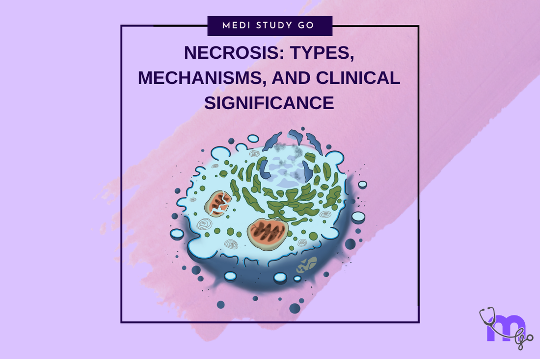
Necrosis: Types, Mechanisms, and Clinical Significance
Medi Study Go
Related Resources
Cell Injury and Adaptation: The Foundation of Pathology
Morphology of Reversible Cell Injury: Key Features and Clinical Examples
Pathogenesis of Reversible Cell Injury: Mechanisms and Outcomes
Pathogenesis of Irreversible Cell Injury: From Damage to Cell Death
Apoptosis: Programmed Cell Death in Health and Disease
Pathological Calcification: Dystrophic vs. Metastatic
Cellular Adaptation: Types, Mechanisms, and Clinical Relevance
Intracellular Accumulations: Lipids, Proteins, Pigments, and More
Cell Injury in Clinical Practice in Dentistry
Key Takeaways
- Necrosis represents uncontrolled cell death resulting from severe injury, characterized by cellular swelling, membrane rupture, and inflammatory response
- Different types of necrosis including coagulative, liquefactive, caseous, and fat necrosis have distinct morphological patterns and clinical implications
- Recognition of necrotic tissue patterns enables appropriate treatment decisions including debridement, antimicrobial therapy, and complication prevention
- Understanding necrosis mechanisms guides clinical management strategies for conditions such as pulpal necrosis, osteonecrosis, and post-surgical complications
- Early recognition and appropriate management of necrotic tissues prevent secondary complications and optimize patient outcomes
Introduction
Necrosis represents the morphological manifestation of irreversible cell death in living tissues, characterized by a spectrum of cellular and tissue changes that result from severe injury exceeding cellular adaptive capacity. For dental professionals, understanding necrosis is essential for recognizing tissue death patterns, implementing appropriate treatment strategies, and preventing secondary complications that can significantly impact patient outcomes.
The process of necrosis involves uncontrolled cellular breakdown following severe injury, distinguished from apoptosis by its disorganized nature, associated inflammatory response, and potential for causing secondary tissue damage. Unlike the controlled process of apoptosis, necrosis results from overwhelming cellular injury that disrupts normal cellular death pathways and leads to cellular content release that can damage surrounding tissues.
Different types of necrosis demonstrate characteristic morphological patterns that provide important diagnostic information and guide appropriate treatment approaches. Recognition of these patterns enables practitioners to assess the extent of tissue damage, predict complications, and implement targeted therapeutic interventions that optimize healing while preventing secondary problems.
In dental practice, necrosis is encountered in various clinical scenarios including pulpal necrosis following deep caries or trauma, osteonecrosis from medication effects or radiation therapy, soft tissue necrosis from ischemia or chemical injury, and post-surgical tissue death from compromised blood supply. Understanding the mechanisms and clinical significance of these conditions is essential for optimal patient management.
Contemporary research continues to elucidate the molecular mechanisms underlying different types of necrosis, providing insights into potential therapeutic targets and prevention strategies. These advances enable more precise diagnostic approaches and targeted interventions that can minimize tissue loss and optimize healing outcomes in conditions involving necrotic tissue.
Table of Contents
Understanding the Pathophysiology of Necrosis What Are the Different Types of Necrosis and Their Characteristics? Coagulative vs. Liquefactive Necrosis in Dental Tissues How Does the Body Respond to Necrotic Tissue? Clinical Management and Treatment Strategies
Understanding the Pathophysiology of Necrosis

Cellular Mechanisms of Necrotic Death
Necrosis occurs when cellular injury exceeds the capacity for adaptation or controlled death pathways, resulting in uncontrolled cellular breakdown and content release. This process involves simultaneous failure of multiple cellular systems including energy production, membrane integrity, and ion homeostasis that creates irreversible cellular damage.
The pathophysiology begins with severe ATP depletion that disrupts essential cellular processes, followed by membrane damage that allows uncontrolled ion flux and cellular swelling. Unlike reversible injury, the extent of damage in necrosis exceeds cellular repair capacity and progresses to inevitable cellular destruction.
Lysosomal enzyme release represents a critical amplifying factor in necrosis, as disruption of these organelles releases digestive enzymes into the cytoplasm, leading to autodigestion of cellular components. This process creates the characteristic morphological changes associated with necrotic death and contributes to secondary tissue damage.
Triggers and Initiating Factors
Hypoxia represents the most common cause of necrosis in dental practice, occurring when tissue oxygen supply becomes inadequate for cellular survival. This can result from vascular compromise, inflammation-induced ischemia, or mechanical compression that prevents adequate blood flow to tissues.
Chemical injury from acids, alkalis, toxins, or inappropriate use of dental materials can cause direct cellular damage that progresses rapidly to necrosis. Understanding these mechanisms enables practitioners to prevent iatrogenic necrosis and manage cases where chemical injury has occurred.
Physical agents including extreme temperatures, radiation, and mechanical trauma can cause immediate cellular death that bypasses normal injury responses and progresses directly to necrosis. Recognition of these causes guides appropriate treatment and prevention strategies.
Inflammatory Response to Necrosis
Necrotic cell death triggers intense inflammatory responses as cellular contents are recognized as foreign by the immune system. This inflammatory response can cause additional tissue damage while serving essential functions in debris removal and healing initiation.
The release of damage-associated molecular patterns (DAMPs) from necrotic cells activates inflammatory cascades that recruit inflammatory cells and initiate tissue repair processes. Understanding this response helps practitioners manage the inflammatory component of necrotic conditions.
Chronic inflammation can develop when necrotic tissue persists or when the inflammatory response becomes dysregulated, leading to ongoing tissue damage and impaired healing. This emphasizes the importance of appropriate necrotic tissue management in clinical practice.
What Are the Different Types of Necrosis and Their Characteristics?
Recognition of different necrosis types provides important diagnostic and therapeutic information, as each type has characteristic morphological features, underlying mechanisms, and clinical implications that guide appropriate management strategies.
Coagulative Necrosis
Coagulative necrosis occurs when cellular proteins become denatured while maintaining basic tissue architecture, resulting in firm, pale tissue with preserved structural outline. This type is most commonly seen in solid organs following ischemic injury and represents the most frequent type of necrosis in dental practice.
The mechanism involves protein denaturation that preserves cellular and tissue structure while eliminating cellular function. This creates characteristic microscopic appearance where cellular outlines remain visible but nuclear detail is lost, indicating cellular death while maintaining tissue organization.
Clinical recognition of coagulative necrosis involves identification of firm, pale, well-demarcated areas of tissue death that maintain their shape and structure. This pattern is commonly observed in pulpal necrosis, osteonecrosis, and ischemic soft tissue injury.
Liquefactive Necrosis
Liquefactive necrosis involves tissue dissolution and liquid formation, typically occurring in tissues with high enzyme content or when bacterial infection is present. This type is characterized by tissue softening and cavity formation that can complicate treatment and healing.
The mechanism involves enzymatic digestion of tissue that liquefies cellular components and creates fluid-filled spaces. This process can be enhanced by bacterial enzymes in infected tissues or by high concentrations of digestive enzymes in certain anatomical locations.
Clinical manifestations include soft, fluctuant masses or cavities filled with necrotic debris that may require drainage and debridement. This pattern is commonly seen in abscesses, infected pulpal necrosis, and certain types of osteomyelitis.
Caseous and Fat Necrosis
Caseous necrosis produces a cheese-like consistency due to incomplete digestion of necrotic tissue, creating a soft, yellowish material that may persist for extended periods. While less common in dental practice, understanding this pattern is important for differential diagnosis.
Fat necrosis occurs specifically in adipose tissue and results from enzymatic breakdown of fat cells, creating characteristic soap formation and potential calcification. This type may be encountered in oral and maxillofacial surgery involving adipose tissue.
The recognition of these specialized necrosis types enables appropriate differential diagnosis and treatment planning, particularly in complex cases involving multiple tissue types or unusual clinical presentations.
Coagulative vs. Liquefactive Necrosis in Dental Tissues
The distinction between coagulative and liquefactive necrosis is particularly important in dental practice as these patterns have different clinical implications and require different management approaches.
Coagulative Necrosis in Dental Practice
Pulpal necrosis typically follows a coagulative pattern in the absence of bacterial infection, resulting in firm, pale pulpal tissue that maintains its basic structure while losing vitality. This pattern allows for predictable endodontic treatment outcomes when managed appropriately.
Periodontal ligament necrosis following trauma or orthodontic forces often demonstrates coagulative patterns that can lead to ankylosis or root resorption if not managed properly. Understanding this mechanism guides appropriate treatment timing and techniques.
Osteonecrosis of the jaws frequently demonstrates coagulative patterns, particularly in medication-related osteonecrosis where bone maintains its structure while losing vitality. This pattern influences surgical management decisions and healing expectations.
Liquefactive Necrosis and Infection
Infected pulpal necrosis progresses to liquefactive patterns as bacterial enzymes digest necrotic tissue, creating conditions that require more extensive debridement and antimicrobial therapy. This pattern indicates more complex treatment requirements and potential complications.
Periodontal abscesses demonstrate liquefactive necrosis patterns that require drainage and debridement in addition to antimicrobial therapy. Understanding this pattern guides appropriate treatment sequencing and technique selection.
Osteomyelitis with liquefactive necrosis requires aggressive surgical debridement and prolonged antimicrobial therapy, as the liquid necrotic material provides an ideal environment for bacterial growth and persistence.
Clinical Decision Making
Recognition of necrosis patterns influences treatment planning decisions including the extent of debridement required, antimicrobial therapy selection, and healing expectations. Coagulative necrosis may require less aggressive intervention while liquefactive necrosis typically requires more extensive treatment.
The presence of infection significantly influences necrosis patterns and treatment requirements, emphasizing the importance of appropriate microbiological assessment and antimicrobial therapy in managing necrotic conditions.
Healing patterns differ between coagulative and liquefactive necrosis, with coagulative necrosis typically showing more predictable healing while liquefactive necrosis may require extended healing periods and closer monitoring for complications.
How Does the Body Respond to Necrotic Tissue?
Acute Inflammatory Response
Necrotic tissue triggers immediate inflammatory responses as cellular contents are recognized as foreign material requiring removal. This response involves vasodilation, increased vascular permeability, and inflammatory cell recruitment that creates the clinical signs of inflammation.
Neutrophil infiltration occurs rapidly as these cells respond to chemotactic signals from necrotic tissue, beginning the process of debris removal through phagocytosis and enzymatic digestion. Understanding this process helps practitioners manage the inflammatory component of necrotic conditions.
The acute inflammatory response serves essential functions in debris removal and healing initiation but can also cause secondary tissue damage if excessive or prolonged. Appropriate management strategies balance the beneficial and harmful aspects of this response.
Chronic Inflammatory Changes
Macrophage activation represents a critical component of the chronic response to necrotic tissue, as these cells are responsible for phagocytosis of large debris particles and coordination of healing responses. Persistent necrotic tissue can lead to chronic macrophage activation and ongoing inflammation.
Granulation tissue formation occurs as part of the healing response to necrotic tissue, involving angiogenesis, fibroblast proliferation, and extracellular matrix deposition that gradually replaces necrotic tissue with healing tissue.
Fibrosis and scarring may result from chronic inflammatory responses to necrotic tissue, particularly when debris removal is incomplete or when healing is complicated by infection or other factors. Understanding these outcomes guides treatment decisions regarding debridement extent and timing.
Healing and Resolution
Debridement processes involve both cellular mechanisms for debris removal and clinical interventions to assist this process. The effectiveness of natural debridement mechanisms influences the need for surgical intervention and the timing of therapeutic procedures.
Tissue regeneration potential varies depending on the tissue type involved and the extent of necrotic damage. Some tissues demonstrate excellent regenerative capacity while others heal primarily through scar formation, influencing treatment goals and expectations.
The balance between debris removal and tissue preservation requires careful clinical judgment, as aggressive debridement may remove viable tissue while inadequate debridement may impair healing and predispose to complications.
Clinical Management and Treatment Strategies
Assessment and Diagnosis
Clinical recognition of necrotic tissue requires understanding of the characteristic signs including tissue discoloration, altered consistency, loss of function, and associated inflammatory responses. These signs enable early recognition and appropriate treatment initiation.
Diagnostic imaging may be helpful in assessing the extent of necrotic tissue, particularly in bone and deep soft tissues where clinical examination may be limited. Understanding the imaging characteristics of different necrosis types guides appropriate diagnostic approaches.
Microbiological assessment is essential when infection is suspected, as the presence of bacteria significantly influences treatment requirements and outcomes. Appropriate specimen collection and culture techniques enable targeted antimicrobial therapy.
Debridement and Surgical Management
Surgical debridement represents the primary treatment for most necrotic conditions, involving removal of non-viable tissue while preserving healthy tissue and optimizing conditions for healing. The extent and technique of debridement depend on the type and location of necrosis.
Conservative debridement approaches may be appropriate for small areas of necrosis or when vital structures are at risk, while aggressive debridement may be necessary for extensive necrosis or when infection is present. Clinical judgment guides appropriate technique selection.
Timing of debridement influences outcomes, with early intervention generally providing better results but requiring careful assessment to distinguish viable from non-viable tissue. Understanding tissue viability markers guides appropriate timing decisions.
Antimicrobial and Supportive Therapy
Antimicrobial therapy is essential when bacterial infection is present or likely, with selection based on likely pathogens, culture results when available, and patient factors including allergies and comorbidities. Understanding common pathogens in different necrotic conditions guides empirical therapy selection.
Supportive care measures including pain management, nutritional support, and optimization of healing conditions can significantly influence outcomes in necrotic conditions. These measures address the systemic effects of necrosis and support natural healing processes.
Monitoring and follow-up protocols ensure appropriate healing progression and early recognition of complications. Understanding expected healing patterns and potential complications guides appropriate monitoring intensity and duration.
Prevention Strategies
Risk factor modification including smoking cessation, diabetes control, and medication management can significantly reduce the risk of developing necrotic conditions. Patient education regarding these factors is essential for prevention.
Iatrogenic prevention involves careful attention to surgical technique, appropriate use of dental materials, and recognition of patient risk factors that predispose to necrosis. Understanding these factors enables preventive strategies during dental procedures.
Early intervention for conditions that predispose to necrosis can prevent progression to tissue death and improve outcomes. Recognition of early warning signs enables timely intervention before irreversible tissue damage occurs.
Conclusion
Understanding necrosis types, mechanisms, and clinical significance provides essential knowledge for recognizing tissue death patterns, implementing appropriate treatment strategies, and optimizing patient outcomes in dental practice. The distinction between different necrosis types guides clinical decision-making regarding debridement extent, antimicrobial therapy, and healing expectations.
Recognition of necrotic tissue patterns enables early intervention that can minimize tissue loss, prevent complications, and optimize healing outcomes. The relationship between necrosis mechanisms and clinical management strategies emphasizes the importance of understanding underlying pathophysiology for effective treatment.
Contemporary approaches to necrosis management continue to evolve with advances in understanding of healing mechanisms, antimicrobial therapy, and tissue preservation techniques. These developments provide opportunities for improved outcomes and reduced morbidity in conditions involving necrotic tissue.
The integration of pathophysiological understanding with clinical practice represents the foundation for optimal management of necrotic conditions in dental medicine. Continued research in this area promises to provide new insights and therapeutic approaches that further improve patient outcomes.