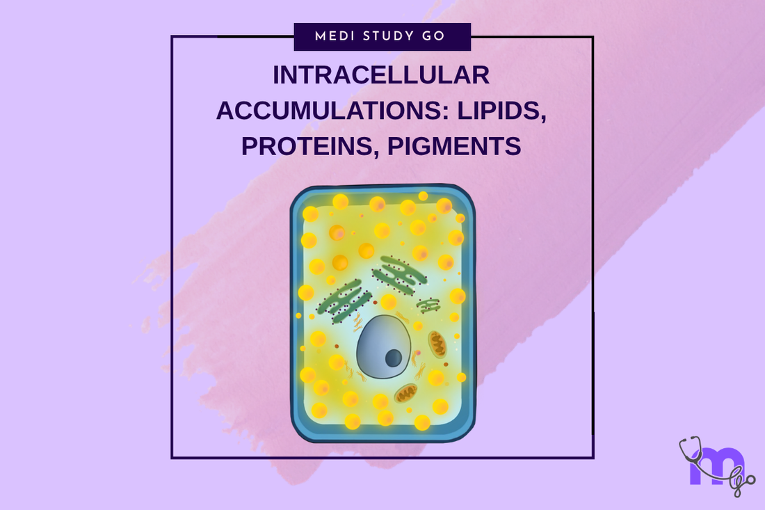
Intracellular Accumulations: Lipids, Proteins, Pigments, and More
Medi Study Go
Related Resources
Cell Injury and Adaptation: The Foundation of Pathology
Morphology of Reversible Cell Injury: Key Features and Clinical Examples
Pathogenesis of Reversible Cell Injury: Mechanisms and Outcomes
Pathogenesis of Irreversible Cell Injury: From Damage to Cell Death
Necrosis: Types, Mechanisms, and Clinical Significance
Apoptosis: Programmed Cell Death in Health and Disease
Pathological Calcification: Dystrophic vs. Metastatic
Cellular Adaptation: Types, Mechanisms, and Clinical Relevance
Cell Injury in Clinical Practice in Dentistry
Key Takeaways
- Intracellular accumulations result from imbalanced cellular metabolism where substance production exceeds elimination or normal degradation pathways become impaired
- Different types of accumulations including lipids, proteins, carbohydrates, and pigments have distinct mechanisms and clinical implications
- Recognition of accumulation patterns helps identify underlying metabolic disorders, genetic defects, or environmental exposures affecting cellular function
- Some accumulations are reversible with appropriate intervention while others represent irreversible storage diseases requiring supportive management
- Understanding accumulation mechanisms guides diagnostic evaluation and enables appropriate treatment of underlying causes when possible
Introduction


Intracellular accumulations represent pathological conditions where normal or abnormal substances accumulate within cells due to imbalanced cellular metabolism, impaired degradation pathways, or overwhelming exposure to foreign materials. For dental professionals, understanding these accumulations is important for recognizing signs of systemic diseases, identifying environmental exposures, and managing conditions that affect oral tissues.
The mechanisms underlying intracellular accumulations involve disruption of normal cellular homeostasis where the rate of substance production or uptake exceeds the cellular capacity for utilization, degradation, or elimination. This imbalance can result from genetic defects, acquired metabolic disorders, environmental exposures, or aging processes that affect cellular function.
Different types of accumulations demonstrate characteristic cellular and tissue changes that provide important diagnostic information. Recognition of these patterns enables identification of underlying causes and guides appropriate management strategies, which may range from elimination of causative factors to supportive care for genetic storage diseases.
In dental practice, intracellular accumulations may be encountered as oral manifestations of systemic diseases, effects of environmental or occupational exposures affecting oral tissues, or age-related changes that influence treatment planning and outcomes. Understanding these conditions enables comprehensive patient care and appropriate medical referral when indicated.
Contemporary research continues to elucidate the molecular mechanisms underlying various accumulation disorders, providing insights into potential therapeutic targets and prevention strategies. These advances offer opportunities for more effective management of accumulation-related conditions and better understanding of their impact on oral health.
Table of Contents
Lipid Accumulations: Fatty Change and Storage Diseases What Causes Protein Accumulations and Cellular Dysfunction? Pigment Accumulations: Endogenous and Exogenous Sources How Do Carbohydrate and Metabolic Accumulations Develop? Clinical Recognition and Management Strategies
Lipid Accumulations: Fatty Change and Storage Diseases
Mechanisms of Lipid Accumulation
Lipid accumulation occurs when cellular lipid uptake exceeds the capacity for lipid oxidation, export, or utilization, leading to triglyceride accumulation in cytoplasmic vacuoles. This process can result from increased lipid availability, impaired oxidation pathways, or defective lipid export mechanisms.
Fatty change represents the most common form of lipid accumulation, typically occurring in liver cells but potentially affecting other tissues including oral structures. The process involves disruption of normal fatty acid metabolism through various mechanisms including hypoxia, toxin exposure, or metabolic disorders.
The cellular mechanisms involve altered fatty acid synthesis, decreased fatty acid oxidation, impaired protein synthesis affecting lipoprotein formation, and defective lipid transport systems. Understanding these mechanisms helps identify potential therapeutic targets and preventive strategies.
Clinical Patterns and Manifestations
Hepatic fatty change represents the most clinically significant form of lipid accumulation, with potential oral manifestations including altered drug metabolism affecting dental treatment responses and increased bleeding tendencies from impaired liver function.
Systemic effects of fatty change can influence oral health through altered immune function, impaired wound healing, and modified responses to medications commonly used in dental practice. Recognition of these effects guides treatment modification and monitoring protocols.
The reversibility of fatty change depends on correction of underlying causes, with most cases showing complete resolution when causative factors are eliminated. This potential for reversibility emphasizes the importance of early recognition and intervention.
Genetic Lipid Storage Diseases
Sphingolipidoses represent inherited disorders of lipid metabolism that can have oral manifestations including gingival enlargement, altered facial features, and developmental abnormalities affecting oral structures. Recognition of these patterns enables early diagnosis and appropriate referral.
Gaucher disease and other lysosomal storage diseases may present with oral findings including delayed tooth eruption, alveolar bone changes, and gingival manifestations that can be among the earliest signs of these conditions.
Understanding genetic lipid storage diseases helps dental professionals recognize patients who may benefit from genetic counseling and enables appropriate modification of dental treatment plans to accommodate systemic complications of these conditions.
What Causes Protein Accumulations and Cellular Dysfunction?
Protein Misfolding and Aggregation
Protein accumulations often result from misfolding of normal proteins that aggregate into insoluble complexes, disrupting normal cellular function and potentially leading to cell death. These processes can result from genetic mutations, environmental stresses, or aging-related changes in protein quality control.
Chaperone protein dysfunction contributes to protein accumulation by impairing the cellular machinery responsible for proper protein folding and preventing aggregation. Understanding these mechanisms provides insights into potential therapeutic approaches for protein accumulation disorders.
The relationship between protein accumulation and cellular stress involves activation of stress response pathways that may initially be protective but can become harmful when overwhelmed by excessive protein accumulation.
Amyloid and Related Deposits
Amyloid deposits consist of misfolded proteins arranged in characteristic beta-pleated sheet configurations that are resistant to normal degradation pathways. These deposits can occur in various tissues and may have oral manifestations in systemic amyloidosis.
Oral manifestations of amyloidosis can include macroglossia, gingival enlargement, and altered tissue texture that may be among the first clinical signs of systemic amyloid disease. Recognition of these signs enables early diagnosis and appropriate medical referral.
The progressive nature of amyloid accumulation and its resistance to normal degradation mechanisms make early recognition and treatment of underlying causes crucial for preventing irreversible tissue damage.
Intracellular Protein Aggregates
Inclusion bodies form when protein accumulation exceeds cellular degradation capacity, creating visible aggregates that can interfere with normal cellular function. These structures may represent cellular attempts to sequester harmful misfolded proteins.
Proteasome dysfunction contributes to protein accumulation by impairing the primary cellular mechanism for degrading damaged or misfolded proteins. Understanding proteasome function provides insights into aging-related protein accumulation.
The cellular consequences of protein accumulation include disrupted organelle function, impaired cellular transport, and activation of cell death pathways when accumulation exceeds cellular tolerance levels.
Clinical Implications and Recognition
Systemic protein accumulation diseases may present with oral manifestations that can aid in early diagnosis, including changes in tongue size, gingival texture, and salivary gland function that reflect underlying protein metabolism disorders.
Age-related protein accumulation contributes to cellular dysfunction and may influence healing responses, treatment outcomes, and susceptibility to oral diseases in elderly patients. Understanding these changes guides age-appropriate treatment modifications.
Environmental factors that promote protein misfolding include oxidative stress, inflammation, and exposure to certain chemicals or medications, emphasizing the importance of comprehensive patient assessment for accumulation risk factors.
Pigment Accumulations: Endogenous and Exogenous Sources
Endogenous Pigment Accumulations
Melanin accumulation can occur in oral tissues due to various stimuli including chronic irritation, hormonal changes, or genetic factors affecting melanocyte activity. Understanding normal and pathological melanin distribution helps distinguish between benign and concerning pigmented lesions.
Lipofuscin represents "wear and tear" pigment that accumulates with aging, resulting from incomplete digestion of cellular components by lysosomes. This accumulation is particularly common in long-lived cells and may influence cellular function over time.
Hemosiderin accumulation occurs following hemorrhage when iron from degraded hemoglobin is stored in tissues, potentially creating characteristic brown discoloration that can persist long after the original bleeding episode.
Exogenous Pigment Deposits
Amalgam tattoos represent common examples of exogenous pigment accumulation in oral tissues, resulting from amalgam particles becoming embedded in tissues during dental procedures. Recognition of these deposits prevents unnecessary concern about malignant pigmentation.
Occupational and environmental pigment exposure can lead to characteristic accumulations in oral tissues, providing clues to occupational hazards or environmental exposures that may require medical evaluation and exposure modification.
Drug-induced pigmentation can occur from various medications including antimalarials, phenothiazines, and other drugs that can deposit in oral tissues, creating characteristic color changes that may be reversible with drug discontinuation.
Clinical Assessment and Management
Pigment accumulation assessment requires careful history taking to distinguish between endogenous and exogenous sources, with different implications for management and prognosis. Understanding pigment sources guides appropriate diagnostic approaches.
Biopsy evaluation may be necessary to definitively characterize pigmented lesions and distinguish between various types of pigment accumulation, particularly when malignancy cannot be excluded based on clinical appearance alone.
Monitoring protocols for pigmented lesions help identify changes that might indicate progression from benign accumulation to pathological processes requiring intervention. Understanding concerning change patterns guides appropriate follow-up intervals.
How Do Carbohydrate and Metabolic Accumulations Develop?
Glycogen Storage Disorders
Glycogen accumulation can occur due to genetic defects in glycogen metabolism, leading to excessive glycogen storage in various tissues including oral structures. These conditions can affect treatment planning and healing responses in dental patients.
Von Gierke disease and other glycogen storage diseases may have oral manifestations including delayed tooth eruption, increased susceptibility to oral infections, and altered wound healing that require treatment modifications.
The management of patients with glycogen storage diseases requires understanding of their metabolic limitations and potential complications that may affect dental treatment safety and outcomes.
Mucopolysaccharide Accumulations
Mucopolysaccharidoses represent inherited disorders of complex carbohydrate metabolism that can have significant oral and facial manifestations including altered facial features, delayed tooth eruption, and gingival changes.
Hurler syndrome and related conditions often present with characteristic oral findings that may be among the first clinical signs of these genetic disorders, emphasizing the importance of recognition by dental professionals.
Understanding mucopolysaccharide accumulation disorders enables appropriate treatment modifications and patient management strategies that accommodate the systemic complications of these conditions.
Metabolic Byproduct Accumulations
Uric acid and other metabolic waste accumulations can affect oral tissues, particularly in patients with renal disease or metabolic disorders affecting waste elimination. Understanding these accumulations guides treatment modification and monitoring.
Calcium accumulations in soft tissues may result from metabolic disorders affecting calcium homeostasis, potentially creating palpable deposits or radiographic changes that require evaluation for underlying systemic disease.
The relationship between metabolic accumulations and systemic disease emphasizes the importance of comprehensive patient assessment and coordination with medical colleagues for optimal management.
Clinical Recognition and Management Strategies
Diagnostic Approaches
Clinical recognition of intracellular accumulations requires systematic evaluation including detailed history taking, physical examination, and appropriate laboratory or imaging studies to characterize the nature and extent of accumulations.
Microscopic examination may be necessary to definitively characterize accumulations and distinguish between different types, particularly when clinical appearance alone cannot provide sufficient diagnostic information.
Specialized staining techniques can help identify specific types of accumulations and provide information about their composition and cellular distribution, guiding appropriate treatment decisions.
Treatment Strategies
Causative factor elimination represents the primary treatment approach for reversible accumulations, emphasizing the importance of identifying and addressing underlying causes when possible.
Supportive care may be appropriate for irreversible accumulations such as genetic storage diseases, focusing on managing complications and optimizing quality of life while accommodating systemic limitations.
Chelation therapy or other specific treatments may be available for certain types of accumulations, particularly those involving toxic metal deposits or specific metabolic disorders.
Prevention and Monitoring
Risk factor identification enables implementation of preventive strategies that minimize exposure to substances that can cause pathological accumulation while optimizing cellular metabolism and elimination pathways.
Regular monitoring protocols help assess accumulation progression and treatment effectiveness while enabling early recognition of complications that may require intervention.
Patient education regarding accumulation disorders helps ensure compliance with treatment recommendations and enables patients to recognize concerning changes that might require medical attention.
Systemic Considerations
Coordination with medical colleagues is essential for patients with systemic accumulation disorders, ensuring comprehensive care that addresses both oral manifestations and underlying systemic disease.
Treatment modifications may be necessary for patients with accumulation disorders, taking into account altered drug metabolism, increased bleeding risk, or other complications that can affect dental treatment safety and outcomes.
Long-term management planning considers the progressive nature of many accumulation disorders and the need for ongoing monitoring and treatment modification as conditions evolve over time.
Conclusion
Understanding intracellular accumulations and their mechanisms provides essential knowledge for recognizing signs of systemic diseases, identifying environmental exposures, and managing conditions that affect oral tissues. The diversity of accumulation types and their various causes emphasizes the importance of comprehensive patient assessment and appropriate diagnostic evaluation.
Recognition of accumulation patterns can provide early clues to underlying systemic diseases, enabling appropriate medical referral and coordinated care that addresses both oral manifestations and systemic conditions. This highlights the important role of dental professionals in comprehensive healthcare.
Clinical management of patients with accumulation disorders requires understanding of their systemic implications and potential effects on dental treatment, emphasizing the need for individualized treatment planning and appropriate monitoring protocols.
Contemporary advances in understanding accumulation mechanisms continue to provide insights into potential therapeutic targets and prevention strategies, offering hope for improved management of these conditions and their oral manifestations.