
Tracheostomy Emergencies in Dentistry: Airway Management and Complications
Medi Study Go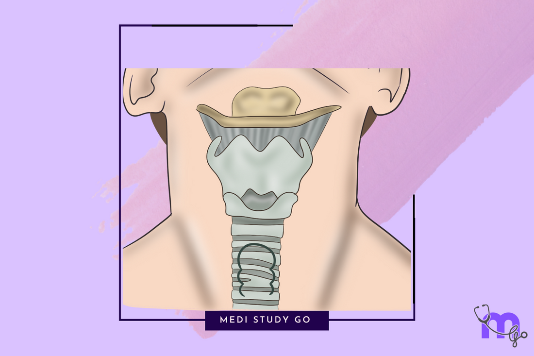
Related Resources:
- Comprehensive Guide to Medical Emergencies in Dentistry
- Cardiopulmonary Resuscitation in Dental Settings
- Syncope Management in Dental Clinics
- Postural Hypotension in Dental Patients
- Anaphylaxis in Dental Practice
- Managing Acute Anginal Attacks
- Diabetic Emergencies in Dentistry
- Anticoagulant Therapy in Dental Patients
- Adrenal Insufficiency Crisis
- Hypertensive Crises in Dental Clinics
- Status Epilepticus and Status Asthmaticus
Introduction
Patients with tracheostomies present unique challenges and considerations in dental settings. A tracheostomy—a surgical opening created in the anterior wall of the trachea to establish an alternative airway—may be present temporarily or permanently in patients with upper airway obstruction, prolonged ventilation needs, or certain head and neck conditions. As the prevalence of community-dwelling individuals with tracheostomies increases, dental professionals are more likely to encounter these patients in their practices. Emergencies involving tracheostomies can develop rapidly and become life-threatening without prompt, appropriate intervention. This comprehensive guide examines the essential aspects of tracheostomy management in dental settings, focusing on emergency recognition, immediate response protocols, and preventive strategies. By understanding tracheostomy anatomy, common complications, and specific airway management techniques, dental professionals can provide safe, effective care for this vulnerable patient population.
Key Takeaways
- Tracheostomy patients require comprehensive pre-treatment assessment and modified dental positioning
- Immediate recognition of tracheostomy emergencies is critical for prompt intervention
- Dental teams should be familiar with common tracheostomy tube types and emergency equipment
- Specialized suctioning techniques are essential for managing secretions and preventing obstruction
- Collaborative care with the patient's medical team optimizes safety and treatment outcomes
Table of Contents
Introduction Understanding Tracheostomies in Dental Patients Pre-treatment Assessment and Planning Common Emergencies and Management Protocols Specialized Equipment and Techniques Preventive Strategies and Risk Reduction Staff Training and Emergency Preparedness Conclusion
Understanding Tracheostomies in Dental Patients
Types and Components of Tracheostomy Tubes
Tracheostomy tubes come in various designs to meet specific patient needs, and dental professionals should be familiar with common configurations:
- Based on material:
- Plastic/PVC tubes: Commonly used for short-term or initial placement
- Metal tubes (silver or stainless steel): Often used for long-term tracheostomies
- Silicone tubes: Provide greater flexibility and comfort for long-term use
- Based on structure:
- Cuffed tubes: Feature an inflatable balloon to seal the airway, preventing aspiration
- Uncuffed tubes: Allow air passage around the tube, permitting speech with digital occlusion
- Fenestrated tubes: Contain an opening on the upper surface allowing airflow to the larynx for speaking
- Inner cannula tubes: Feature a removable inner tube for easy cleaning without complete tube removal
- Key components:
- Outer cannula: Main tube inserted into the trachea
- Inner cannula: Removable liner that can be cleaned or replaced
- Obturator: Guide used during insertion (removed after placement)
- Flange/neck plate: External component that secures the tube to the neck
- Cuff and pilot balloon: Inflation system for sealing the airway when present
Understanding these variations helps dental professionals anticipate potential complications and maintenance needs during treatment.
Physiological Implications for Dental Treatment
Tracheostomy presence significantly alters respiratory physiology with several implications for dental care:
- Airway dynamics:
- Bypassing of upper airway natural humidification and filtering
- Altered mucociliary clearance mechanisms
- Direct exposure of lower airways to environmental pathogens
- Reduced ability to generate positive pressure (affecting coughing efficacy)
- Communication challenges:
- Impaired or absent vocalization with standard tracheostomy tubes
- Reliance on alternative communication methods
- Need for modified informed consent processes
- Psychosocial considerations:
- Potential anxiety about airway management in the dental setting
- Self-consciousness about tracheostomy appearance
- Concerns about secretion management during procedures
These physiological and psychosocial factors necessitate modifications to standard dental protocols, particularly regarding patient positioning, suction access, and communication strategies.
Pre-treatment Assessment and Planning
Comprehensive History and Evaluation
Thorough pre-treatment assessment for tracheostomy patients includes:
- Medical history specifics:
- Underlying condition necessitating tracheostomy
- Duration of tracheostomy presence
- Anticipated permanence or plans for decannulation
- Recent respiratory infections or complications
- Current ventilation requirements (if any)
- Oxygen dependency assessment
- Tracheostomy-specific information:
- Tube type, size, and manufacturer
- Cuff status (inflated/deflated during normal activities)
- Cleaning routine and frequency
- Recent tube changes and any complications
- Suctioning frequency and typical secretion characteristics
- Speech capability and preferred communication methods
- Emergency information:
- Contact details for managing physician/team
- Patient-specific emergency protocols
- Location of spare tubes or emergency equipment
- Previous complications and their management
This detailed assessment allows for customized treatment planning and emergency preparation. Documentation should be comprehensive, easily accessible, and regularly updated.
Treatment Modifications and Positioning Considerations
Dental treatment for tracheostomy patients requires specific modifications:
- Appointment scheduling:
- Longer appointments to accommodate positioning and suctioning needs
- Morning scheduling when secretions are typically minimal
- Avoiding scheduling immediately after tracheostomy tube changes
- Positioning protocol:
- Semi-upright positioning (30-45° incline) to reduce aspiration risk
- Neck extension minimization to prevent tube displacement
- Access maintenance for emergency equipment
- Regular position adjustments to facilitate secretion drainage
- Infection control enhancements:
- HEPA filtration in treatment room if available
- Minimized aerosol-generating procedures
- Enhanced PPE including face shields
- Preprocedural antimicrobial mouth rinses
- Monitoring requirements:
- Continuous visual observation of respiratory status
- Pulse oximetry throughout treatment
- Regular assessment of tube position and patency
- Vigilance for signs of respiratory distress
These modifications should be documented in the patient's treatment plan and communicated to all team members involved in care delivery.
Common Emergencies and Management Protocols
Tube Obstruction and Dislodgement
Tube obstruction represents the most common and potentially life-threatening tracheostomy emergency:
- Obstruction causes:
- Mucus plugging (most common)
- Blood clots following procedures
- Aspiration of dental materials
- Tube positioning against tracheal wall
- Foreign body introduction
- Recognition of obstruction:
- Increased respiratory rate and effort
- Decreased oxygen saturation
- Anxiety and distress
- Inability to speak or cough effectively
- Cyanosis (late sign)
- Immediate management protocol:
- Stop dental procedure immediately
- Position patient upright
- Attempt suctioning through tube using appropriate catheter
- If unsuccessful, remove inner cannula (if present) for cleaning
- Administer supplemental oxygen alongside tracheostomy
- If obstruction persists, replace entire tube if trained or activate emergency services
Tube dislodgement requires equally urgent intervention:
- Types of dislodgement:
- Partial: Tube partially removed but still providing some airway access
- Complete: Tube entirely removed from stoma
- False passage: Tube displaced into tissues rather than airway
- Management of dislodgement:
- For established tracheostomy (>7 days old):
- Attempt reinsertion if trained and replacement tube available
- Use lubrication and gentle technique following anatomical curve
- Confirm placement with end-tidal CO₂ if available
- For recent tracheostomy (<7 days old):
- Do not attempt reinsertion (risk of false passage)
- Maintain stoma patency with tracheal dilators if available
- Cover stoma with oxygen delivery
- Activate emergency services immediately
- For established tracheostomy (>7 days old):
Dental teams should have algorithms for these emergencies visibly posted in treatment areas.
How to manage tracheostomy-related bleeding during oral surgery?
Management of tracheostomy-related bleeding during oral surgery requires a systematic approach:
- Immediate actions:
- Discontinue the procedure and position patient upright
- Identify bleeding source (stoma site, within trachea, or oral surgical site)
- Apply direct pressure with sterile gauze for external bleeding
- Implement gentle suctioning to maintain airway while clearing blood
- Avoid suctioning with excessive force which may traumatize tissues
- Intervention strategies:
- For minor bleeding:
- Apply topical hemostatic agents appropriate for the bleeding site
- Consider vasoconstrictor-containing local anesthetics for stoma-site bleeding
- Use cold saline irrigation to promote vasoconstriction
- For moderate to severe bleeding:
- Replace standard tracheostomy tube with cuffed tube if bleeding originates above cuff level
- Inflate cuff to create tamponade effect
- Consider topical application of 1:1000 epinephrine on gauze (with caution)
- Activate emergency services if bleeding persists
- For minor bleeding:
- Stabilization measures:
- Monitor oxygen saturation continuously
- Maintain intravenous access if established
- Assess for signs of respiratory compromise
- Consider prophylactic antibiotics if bleeding creates aspiration risk
Prevention remains key, with careful preoperative assessment of hemorrhage risk and surgical planning to avoid structures near the tracheostomy site.
Respiratory Infections and Aspiration
Tracheostomy patients have increased susceptibility to respiratory complications during dental treatment:
- Infection risk factors:
- Direct lower airway access bypassing natural defenses
- Increased mucus production and stasis
- Proximity of dental pathogens to tracheostomy site
- Compromised cough reflex in many patients
- Prevention strategies:
- Pretreatment prophylactic antibiotics (consult with physician)
- Enhanced oral hygiene protocols before invasive procedures
- Use of chlorhexidine-soaked gauze around stoma during treatment
- Minimizing aerosol-generating procedures
- Aspiration management:
- Immediate upright positioning
- Suctioning via tracheostomy tube
- Oxygen supplementation
- Assessment for respiratory distress
- Emergency service activation for significant aspiration
Preventive measures should be integrated into the treatment plan, with emergency protocols readily available if complications arise despite precautions.
Specialized Equipment and Techniques
Suctioning Procedures and Equipment
Proper suctioning is essential for tracheostomy care and emergency management:
- Suctioning equipment requirements:
- Portable or wall suction unit capable of 100-150 mmHg pressure
- Appropriate suction catheters (typically 10-14 Fr for adults)
- Sterile saline or water for irrigation
- Clean gloves and eye protection
- Supplemental oxygen
- Suctioning technique:
- Pre-oxygenate patient if possible
- Use sterile technique for catheter handling
- Insert catheter without suction until resistance felt
- Apply suction only during withdrawal
- Limit suctioning duration to <10 seconds
- Allow respiratory recovery between attempts
- Maximum 2-3 passes per suctioning session
- Specialized considerations during dental procedures:
- Position suction equipment within immediate reach
- Designate a specific team member for airway monitoring and suctioning
- Consider prophylactic suctioning before and after aerosol-generating procedures
- Ensure dental suction does not interfere with access to tracheostomy
Proper suctioning technique prevents trauma while effectively clearing secretions and maintaining airway patency during dental procedures.
Emergency Airway Management Tools
Dental practices treating tracheostomy patients should maintain specific emergency equipment:
- Essential emergency items:
- Tracheostomy dilators
- Appropriately sized replacement tubes (ideally patient's specific type)
- Bag-valve-mask device with tracheostomy adapter
- Water-soluble lubricant
- Suction equipment with various catheter sizes
- Oxygen delivery system with tracheostomy attachments
- Supplementary equipment:
- End-tidal CO₂ monitors
- Pulse oximeter
- Handheld nebulizer for saline delivery
- Heat and moisture exchangers
- Sterile gauze and cleaning supplies
- Patient-specific items (requested from patient):
- Spare tracheostomy tube of current size
- One size smaller tube for emergency use
- Patient-specific suctioning catheters
- Familiar communication devices
This equipment should be organized in a dedicated tracheostomy emergency kit, regularly inspected, and located in an easily accessible area of the treatment room.
Preventive Strategies and Risk Reduction
Collaborative Care Approaches
Optimal care for tracheostomy patients requires interdisciplinary collaboration:
- Communication with medical providers:
- Obtaining current tracheostomy care plan
- Confirming appropriate dental modifications
- Discussing procedure-specific risks
- Establishing emergency protocols
- Determining need for prophylactic medications
- Engaging specialized providers:
- Speech-language pathologists for communication strategies
- Respiratory therapists for airway management guidance
- Home care nurses familiar with patient's routine care
- Otolaryngologists for procedure-specific precautions
- Documentation and information sharing:
- Obtaining and maintaining current medical reports
- Sharing dental treatment plans with medical team
- Documenting all communications in patient record
- Developing unified emergency action plans
A team-based approach significantly reduces emergency risks while enhancing overall care quality and coordination.
Environmental and Procedural Safeguards
The dental environment can be modified to enhance safety for tracheostomy patients:
- Treatment room adaptations:
- Optimized lighting for tracheostomy visualization
- Increased space for equipment and emergency access
- Minimized particulate matter (HEPA filtration when possible)
- Enhanced infection control protocols
- Procedural modifications:
- Alternative materials and techniques to minimize aspiration risk
- Modified radiographic approaches to accommodate tube
- Rubber dam use whenever possible
- Anti-aspiration positioning and draping
- Staff preparation:
- Assigning specific roles for routine and emergency care
- Pre-procedure briefings on patient-specific considerations
- Ensuring familiarity with all necessary equipment
- Confirming emergency protocol understanding
These safeguards should be systematically implemented for all tracheostomy patients, with additional measures based on individual risk assessment.
Staff Training and Emergency Preparedness
Education and Simulation
Effective tracheostomy emergency management requires dedicated training:
- Essential knowledge components:
- Tracheostomy anatomy and physiology
- Common tube types and components
- Basic troubleshooting techniques
- Recognition of emergency situations
- Proper suctioning methodology
- Skill development approaches:
- Hands-on practice with various tracheostomy tubes
- Suctioning technique demonstration and practice
- Tube change observation and supervised practice
- Use of simulation mannequins when available
- Emergency response training:
- Regular simulation of common emergencies
- Role assignment and team communication practice
- Timed response drills with realistic scenarios
- Post-drill debriefing and improvement planning
Training should be repeated regularly with documentation of staff competencies and areas for continued development.
Protocol Development and Implementation
Standardized protocols enhance emergency preparedness:
- Written protocol components:
- Step-by-step emergency response algorithms
- Equipment checklists and maintenance schedules
- Communication pathways within and outside practice
- Documentation requirements for incidents
- Quality improvement mechanisms
- Implementation strategies:
- Visible posting of essential protocols in treatment areas
- Regular review during staff meetings
- Protocol integration into electronic health records
- Patient-specific protocol customization
- Annual review and updating based on current guidelines
- Quality assurance measures:
- Incident reporting system
- Regular equipment function testing
- Mock emergency evaluations
- Feedback mechanisms for improvement
These protocols should be living documents, regularly reviewed and refined based on experience, current evidence, and evolving patient needs.
Conclusion
Tracheostomy patients in dental settings present unique challenges that require specialized knowledge, preparation, and protocols. By understanding the anatomical and physiological implications of tracheostomies, dental professionals can anticipate potential complications and implement preventive strategies. Comprehensive pre-treatment assessment, appropriate modifications to dental procedures, and collaborative care with medical providers form the foundation of safe management.
The ability to promptly recognize and effectively manage tracheostomy emergencies—particularly tube obstruction, dislodgement, and bleeding—can be life-saving. Through dedicated staff training, simulation exercises, and proper equipment preparation, dental practices can develop confidence and competence in caring for this vulnerable patient population.
As the number of community-dwelling individuals with tracheostomies continues to increase, dental professionals have both the opportunity and responsibility to develop expertise in their management. By implementing the strategies outlined in this guide, dental practices can ensure they provide safe, effective, and compassionate care to tracheostomy patients while being prepared for any emergencies that may arise