
Syncope Management in Dental Clinics: Causes, Prevention, and Immediate Response Strategies
Medi Study Go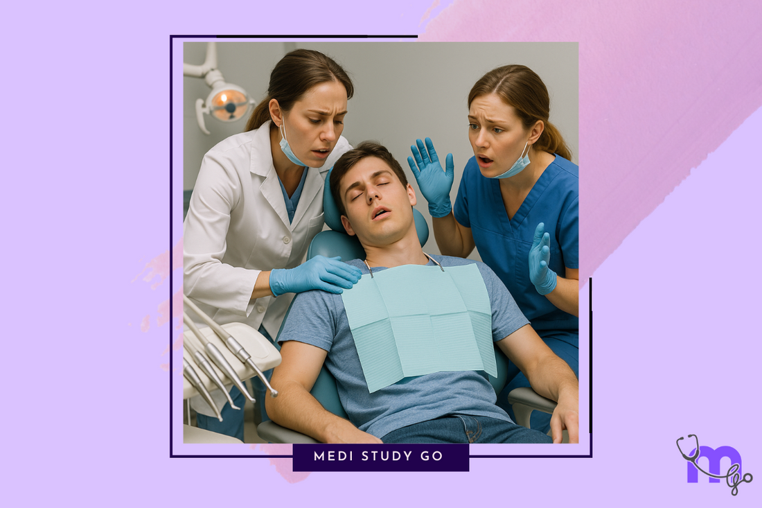
Related Resources:
- Comprehensive Guide to Medical Emergencies in Dentistry
- Cardiopulmonary Resuscitation in Dental Settings
- Postural Hypotension in Dental Patients
- Tracheostomy Emergencies in Dentistry
- Anaphylaxis in Dental Practice
- Managing Acute Anginal Attacks
- Diabetic Emergencies in Dentistry
- Anticoagulant Therapy in Dental Patients
- Adrenal Insufficiency Crisis
- Hypertensive Crises in Dental Clinics
- Status Epilepticus and Status Asthmaticus
Introduction
Syncope, commonly known as fainting, is the most frequent medical emergency encountered in dental practices. Characterized by a temporary loss of consciousness due to inadequate cerebral blood flow, syncope accounts for approximately 50% of all medical emergencies in dental settings. The dental environment creates perfect storm of syncope-inducing factors: patient anxiety, pain, prolonged sitting in the dental chair, and the sight of dental instruments or blood. This comprehensive guide explores the pathophysiology, recognition, management, and prevention of syncope in dental clinics, providing dental professionals with essential knowledge to handle these incidents confidently and effectively.
Key Takeaways
- Syncope is primarily caused by cerebral hypoperfusion and is often preceded by recognizable prodromal symptoms
- The Trendelenburg position (supine with legs elevated) is the cornerstone of immediate management
- Most syncope episodes resolve quickly with proper positioning and supportive care
- Differentiating syncope from more serious conditions like hypoglycemia or cardiac events is crucial
- Preventive strategies include patient screening, anxiety management, and proper positioning
Table of Contents
Introduction Pathophysiology of Syncope Recognition and Assessment Immediate Management Protocols Differential Diagnosis Prevention Strategies Documentation and Follow-up Conclusion
Pathophysiology of Syncope
Understanding the Mechanisms
Syncope results from a temporary reduction in cerebral blood flow, typically caused by one of three primary mechanisms: vasovagal (neurally mediated), orthostatic (postural), or cardiac. In dental settings, vasovagal syncope predominates, triggered by emotional stress that activates the parasympathetic nervous system while inhibiting sympathetic tone. This leads to bradycardia, vasodilation, hypotension, and subsequently, reduced cerebral perfusion.
The autonomic response occurs in stages, beginning with a sympathetic surge (fight-or-flight response) characterized by tachycardia and increased blood pressure. This is followed by parasympathetic predominance, causing bradycardia, peripheral vasodilation, and venous pooling. When cerebral blood flow drops below the critical threshold (approximately 30% below normal), consciousness is lost as the brain attempts to restore normal supine positioning.
Risk Factors and Predisposing Conditions
Several factors increase syncope risk in dental settings:
- Patient-related factors:
- Young adults (15-35 years)
- History of previous syncope
- Anxiety disorders
- Fasting before appointments
- Medication effects (antihypertensives, diuretics)
- Dehydration
- Anemia
- Procedure-related factors:
- Prolonged sitting in semi-supine position
- Pain or painful procedures
- Sight of blood or instruments
- Local anesthetic injections
- Hot, humid environment in treatment room
Identification of these risk factors during pre-treatment assessment allows for preventive measures to be implemented before clinical procedures begin.
Recognition and Assessment
Prodromal Signs and Symptoms
The ability to recognize prodromal symptoms (presyncope) allows for intervention before complete loss of consciousness. These warning signs typically include:
- Subjective symptoms:
- Lightheadedness or dizziness
- Nausea or feeling of warmth
- Visual disturbances (blurring, tunnel vision)
- Tinnitus or hearing changes
- Sense of impending doom
- Observable signs:
- Pallor (often first visible sign)
- Sweating/diaphoresis
- Yawning or sighing respirations
- Pupillary dilation
- Confused or slurred speech
The prodromal phase may last from seconds to several minutes. Prompt recognition during this phase allows for immediate intervention that may prevent progression to complete syncope.
Clinical Assessment and Monitoring
When syncope is suspected, rapid assessment should include:
- Consciousness level: Assess responsiveness and orientation
- Vital signs: Check pulse rate, rhythm, and blood pressure
- Respiratory status: Observe rate, depth, and pattern
- Skin signs: Note color, temperature, and moisture
During a typical vasovagal episode, the pulse is usually slow but regular, blood pressure is low, and the skin is pale, cool, and diaphoretic. This clinical picture helps differentiate vasovagal syncope from other causes of decreased consciousness, such as hypoglycemia or cardiac arrhythmias.
Immediate Management Protocols
Position and Basic Support
The cornerstone of syncope management is proper positioning:
- For prodromal symptoms:
- Immediately discontinue dental treatment
- Position patient in Trendelenburg position (supine with legs elevated approximately 15-20°)
- Ensure open airway and loosen any restrictive clothing
- Maintain verbal reassurance and encourage deep breathing
- For complete loss of consciousness:
- Ensure patient safety to prevent falls or injury
- Place in supine position with legs elevated
- Confirm airway patency and breathing
- Monitor vital signs continuously
- Provide supplemental oxygen if available
The Trendelenburg position increases venous return to the heart, improving cardiac output and cerebral perfusion. Recovery typically begins within 15-30 seconds of proper positioning, with full consciousness usually returning within 1-2 minutes.
Pharmacological Interventions and Advanced Measures
Pharmacological interventions are rarely necessary for simple vasovagal syncope but may include:
- Ammonia inhalants (smelling salts): May stimulate respiration and consciousness, but use is controversial
- Atropine: Reserved for persistent symptomatic bradycardia (HR < 40 bpm)
- IV fluids: For patients with suspected hypovolemia or prolonged episodes
If syncope does not resolve promptly with basic measures, or if atypical features are present, activation of emergency medical services is warranted. While awaiting advanced care, continue to monitor vital signs and maintain proper positioning.
Differential Diagnosis
Distinguishing Syncope from Other Conditions
Several conditions may mimic syncope and require different management approaches:
- Hypoglycemia: Characterized by gradual onset, confusion, diaphoresis, and tachycardia before loss of consciousness. Blood glucose testing can confirm diagnosis.
- Seizure: Distinguished by tonic-clonic movements, tongue biting, urinary incontinence, and prolonged post-ictal confusion.
- Cardiac events: Often preceded by chest pain, palpitations, or shortness of breath; may be accompanied by arrhythmias.
- Stroke: Presents with focal neurological deficits, asymmetrical symptoms, and prolonged unconsciousness.
- Drug-related reactions: Consider medication effects or allergic reactions, especially with recent drug administration.
The key distinguishing features of vasovagal syncope include its typical triggers, prodromal symptoms, brief duration, and rapid recovery without neurological deficits.
When to Activate Emergency Services
Emergency medical services should be activated if:
- Consciousness is not regained within 2 minutes
- Recurrent episodes occur during the same appointment
- Abnormal vital signs persist after recovery
- The patient has known cardiovascular disease
- The episode was not preceded by typical prodromal symptoms
- Seizure activity, chest pain, or focal neurological signs are present
When in doubt, it is better to activate emergency services, as some serious conditions may initially present similar to simple syncope.
Prevention Strategies
Pretreatment Assessment and Risk Reduction
Effective prevention begins with thorough pretreatment screening:
- Medical history review:
- Previous syncope episodes
- Cardiac or neurological conditions
- Current medications
- Fasting status and hydration
- Psychosocial assessment:
- Anxiety levels and dental phobias
- Previous traumatic dental experiences
- Current stress levels
- Risk-reduction strategies:
- Schedule anxious patients for morning appointments
- Encourage normal eating before appointments
- Ensure adequate hydration
- Consider prophylactic positioning (semi-reclined rather than upright)
For patients with multiple risk factors or previous syncope episodes, consider incremental approach to treatment with shorter appointments.
Anxiety Management and Patient Positioning
Anxiety management techniques significantly reduce syncope risk:
- Psychological approaches:
- Systematic desensitization
- Distraction techniques (music, television)
- Guided imagery or relaxation exercises
- Effective communication and explanation of procedures
- Pharmacological approaches:
- Oral anxiolytics (benzodiazepines) for severe dental anxiety
- Nitrous oxide-oxygen sedation
- Conscious sedation for high-risk patients
- Optimal positioning:
- Gradual position changes to prevent orthostatic hypotension
- Slight head elevation (10-15°) during lengthy procedures
- Periodic changes in chair position during long appointments
For patients with recurrent vasovagal episodes, applied muscle tension techniques can be taught, which involve isometric contraction of large muscle groups to increase peripheral resistance and cardiac output.
Documentation and Follow-up
Recording and Reporting Incidents
Thorough documentation of syncope episodes is essential:
- Detailed incident report including:
- Time and circumstances of the event
- Prodromal symptoms and duration
- Loss of consciousness duration
- Vital signs during and after the episode
- Interventions performed
- Patient's response to interventions
- Resolution and final disposition
- Communication with patient's primary care provider:
- Formal referral letter for first-time syncope
- Details of the episode and management
- Recommendations for follow-up evaluation
Complete documentation serves both clinical and legal purposes, demonstrating appropriate care and facilitating future risk assessment.
Post-incident Patient Education
Patient education after a syncope episode should include:
- Explanation of the event and its causes
- Preventive strategies for future dental visits
- Recognition of prodromal symptoms
- Importance of medical follow-up if indicated
- Guidance on safe transportation home (patients should not drive after syncope)
Written post-care instructions should be provided, with recommendation to avoid driving or operating machinery for at least 24 hours after a syncope episode.
Conclusion
Syncope in dental settings, while alarming, can be effectively managed with proper preparation and response. By understanding the underlying pathophysiology, recognizing prodromal signs, implementing immediate management protocols, and developing preventive strategies, dental professionals can minimize both the occurrence and consequences of syncope episodes.
The key to successful management lies in early recognition and prompt intervention, particularly with proper positioning. Most vasovagal syncope episodes resolve quickly and completely with basic measures, allowing dental treatment to be rescheduled or, in some cases, continued after full recovery.
By integrating syncope management into comprehensive emergency protocols and providing regular staff training, dental practices can ensure patient safety while fostering confidence in handling this common emergency situation.