Implant Retention and Stability: Effectiveness Without Pulling Out
Medi Study Go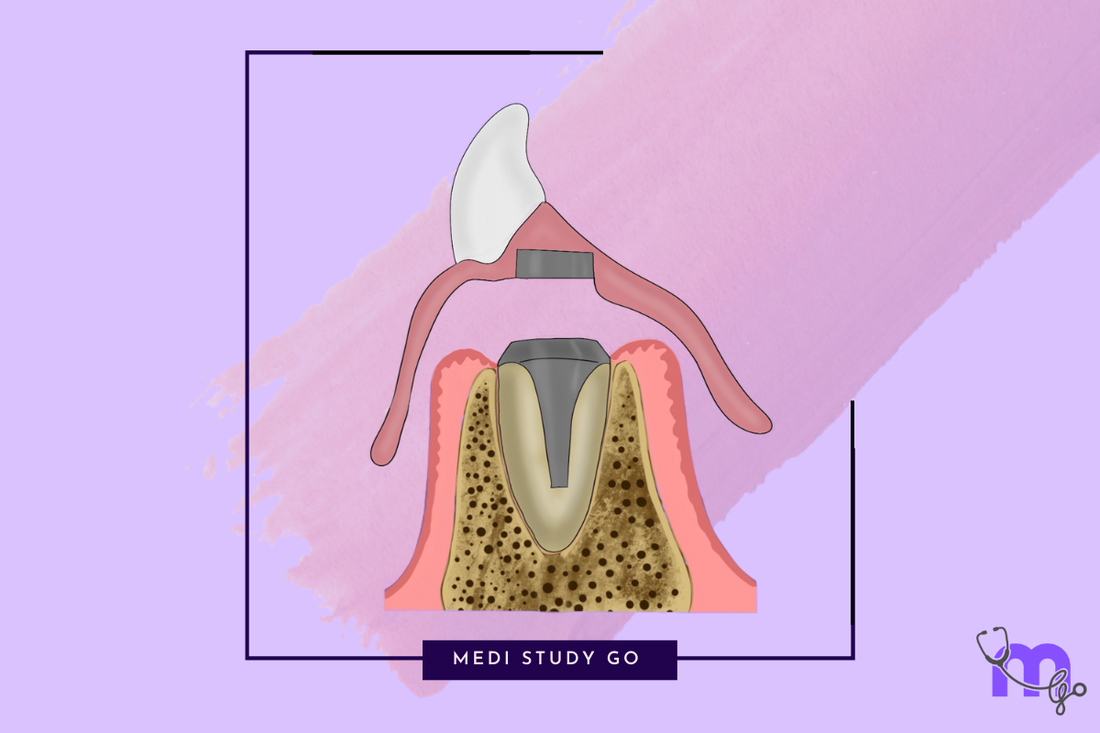
Related Resources:
- Understanding Maxillofacial Prosthodontics: Definition and Scope
- Maxillofacial Prosthodontics and Implantology: The Synergistic Relationship
- Implant Placement and Positioning: Where Does the Implant Go?
- Advanced Techniques in Maxillofacial Prosthodontics
Introduction: The Cornerstone of Implant Success
The effectiveness of implants without pulling out represents perhaps the most critical determinant of long-term success in implant-supported maxillofacial rehabilitation. While proper placement creates the foundation, it is the ongoing stability and retention of both the implant within bone and the prosthesis upon the implant that ultimately delivers functional and aesthetic outcomes for patients.
For students preparing for NEET MDS examinations, understanding the biological and mechanical factors influencing implant stability provides essential knowledge for both theoretical questions and clinical practice. For practicing clinicians, this knowledge guides critical decisions throughout the treatment process, from initial assessment to long-term maintenance.
"The most brilliantly designed prosthesis is only as good as the stability of the implants supporting it. Understanding the science of implant retention transforms clinical outcomes from merely functional to truly life-changing."
This article explores the complex factors that determine implant stability in maxillofacial prosthodontics, from initial placement through long-term function, with special consideration for the unique challenges presented by maxillofacial defects.

Understanding Implant Stability: A Two-Phase Process
Implant stability occurs in two distinct but interrelated phases that must be understood separately.
Primary Stability: The Mechanical Foundation
Primary stability refers to the initial mechanical engagement achieved at implant placement:
- Definition: The absence of mobility in the implant-bone interface immediately after placement
- Mechanical Nature: Achieved through physical interlocking of the implant with surrounding bone
- Importance: Critical for preventing micromotion during the healing phase
- Measurement: Often quantified through insertion torque values or resonance frequency analysis
- Determinants: Bone density, implant design, surgical technique, and site preparation
Secondary Stability: The Biological Connection
Secondary stability refers to the biological stability achieved through osseointegration:
- Definition: The direct structural and functional connection between living bone and the implant surface
- Biological Nature: Achieved through bone remodeling and new bone formation
- Timeline: Typically developing over 2-6 months, depending on various factors
- Measurement: Resonance frequency analysis, absence of radiolucency, clinical stability
- Determinants: Primary stability, bone healing capacity, implant surface, patient factors
The Stability Dip Phenomenon
Understanding the relationship between these two stability phases is crucial:
- Initial Stability Decline: As remodeling begins, some primary stability is lost
- Transition Period: A critical window when stability may temporarily decrease
- Secondary Stability Development: Gradually increases as osseointegration progresses
- Net Stability: Eventually exceeds initial primary stability through biological integration
Factors Affecting Implant Stability in Maxillofacial Applications
Multiple variables influence whether an implant will remain stable throughout function.
Bone-Related Factors
The quality and quantity of available bone significantly impact stability:
-
Bone Density Classification:
- Type I: Dense cortical bone (highest primary stability)
- Type II: Thick cortical with dense trabecular core
- Type III: Thin cortical with dense trabecular bone
- Type IV: Thin cortical with sparse trabecular bone (lowest primary stability)
-
Bone Volume Considerations:
- Height: Determines potential implant length and engagement
- Width: Affects implant diameter selection and cortical engagement
- Three-Dimensional Architecture: Especially important in reconstructed areas
-
Bone Healing Capacity:
- Vascular Supply: Critical for nutrient delivery and cellular activity
- Cellular Activity: Osteoblast and osteoclast function
- Remodeling Potential: Ability to respond to mechanical loading
Implant Design Factors
The physical characteristics of the implant significantly influence stability:
-
Macro Design Features:
- Thread Design: Deeper, more widely spaced threads generally increase primary stability
- Implant Shape: Tapered designs often achieve higher primary stability than parallel-walled
- Diameter: Wider implants typically provide greater initial stability
- Length: Contributes to overall surface area and potential bone engagement
-
Surface Characteristics:
- Roughness: Moderately rough surfaces (1-2μm) generally optimize osseointegration
- Surface Treatments: Sandblasting, acid-etching, oxidation, or hydroxyapatite coating
- Hydrophilicity: More hydrophilic surfaces may accelerate early healing
- Bioactive Coatings: Growth factors or other biological mediators
Surgical Technique Factors
How the implant is placed significantly affects its stability:
-
Site Preparation:
- Drilling Protocol: Standard, undersized, or oversized preparation
- Drilling Speed: Impact on heat generation and bone trauma
- Cooling: Adequate irrigation to prevent thermal necrosis
- Incremental Approaches: Gradual site expansion in soft bone
-
Insertion Technique:
- Insertion Torque: Optimal range typically 30-45 Ncm
- Thread Engagement: Maximizing contact with available bone
- Final Positioning: Subcrestal, crestal, or supracrestal placement
- Immediate Protocols: Special considerations for immediate placement
Patient-Related Factors
Individual patient characteristics significantly influence implant stability:
-
Systemic Health:
- Metabolic Disorders: Diabetes, osteoporosis
- Immune Function: Autoimmune conditions, immunosuppression
- Nutritional Status: Vitamin D, calcium, protein intake
-
Local Factors:
- Previous Radiation: Significantly compromises bone healing
- Soft Tissue Quality: Affects biological seal and protection
- Parafunctional Habits: Bruxism, clenching
- Oral Hygiene: Impact on peri-implant health
-
Behavioral Factors:
- Smoking: Significantly reduces success rates
- Alcohol Consumption: May impact healing and maintenance
- Compliance: With follow-up and maintenance protocols
Special Considerations in Maxillofacial Defects
Maxillofacial defect cases present unique challenges for implant stability.
Post-Surgical and Post-Radiation Considerations
Patients with maxillofacial defects often have compromised tissues:
-
Altered Anatomy:
- Disrupted cortical boundaries
- Changed biomechanical load patterns
- Modified soft tissue relationships
-
Radiation Effects:
- Hypocellularity
- Hypovascularity
- Hypoxia
- Increased risk of osteoradionecrosis
-
Reconstructed Sites:
- Variable bone density in grafts
- Modified blood supply
- Different healing patterns
- Potential for resorption
Modified Protocols for Compromised Sites
Several strategies can enhance stability in challenging maxillofacial cases:
-
Extended Healing Periods:
- Longer osseointegration time before loading
- Gradual loading protocols
- Progressive prosthetic complexity
-
Enhanced Primary Stability Techniques:
- Bicortical engagement when possible
- Undersized preparation in soft bone
- Strategic implant distribution
-
Specialized Implant Selection:
- Surface treatments optimized for compromised bone
- Wider diameter implants where bone allows
- Designs maximizing available bone contact
-
Adjunctive Procedures:
- Hyperbaric oxygen therapy in irradiated bone
- Growth factor applications
- Bone quality enhancement techniques
Assessment Methods for Implant Stability
Various clinical and technological approaches help evaluate implant stability.
Clinical Assessment Techniques
Simple clinical methods provide valuable information:
-
Percussion Testing:
- Sharp, clear tone suggests good integration
- Dull sound may indicate fibrous encapsulation
- Limitations in sensitivity and specificity
-
Mobility Testing:
- Applied pressure with instrument handles
- Contraindicated once osseointegration is established
- Primarily useful for detecting gross failure
-
Torque Testing:
- Reverse torque application (typically 20 Ncm)
- Destructive if applied during integration
- Used selectively in specific clinical situations
Technological Assessment Tools
Advanced technology offers more objective measurements:
-
Resonance Frequency Analysis (RFA):
- Measures implant stability quotient (ISQ)
- Non-invasive and repeatable
- Provides quantitative values (1-100)
- Higher values indicate greater stability
- Useful for tracking stability over time
-
Insertion Torque Measurement:
- Quantifies resistance during placement
- Predictive of primary stability
- Typically measured in Ncm
- Target values usually 30-45 Ncm
- Limited to installation phase only
-
Damping Capacity Assessment:
- Measures energy dissipation upon impact
- Reflects bone density around implant
- Quantifies stability without applying torque
-
Radiographic Evaluation:
- Assesses bone-to-implant contact
- Identifies radiolucencies
- Limited by two-dimensional nature of standard radiographs
- Enhanced by 3D imaging techniques
Prosthetic Retention Mechanisms in Maxillofacial Applications

Once implant stability is established, various systems connect the prosthesis to the implants.
Fixed Prosthesis Retention
For permanently attached restorations:
-
Screw-Retained Systems:
- Direct screw connection to implant or abutment
- Excellent retrievability
- No cement-related complications
- Requires precise implant positioning
- Access holes may compromise aesthetics
-
Cement-Retained Systems:
- Conventional cementation to abutments
- Superior aesthetics without access holes
- Simplified passive fit
- Challenges with excess cement removal
- Limited retrievability
-
Combination Approaches:
- Screw-retained frameworks with cemented superstructures
- Combines advantages of both systems
- Especially valuable in extensive reconstructions
Removable Prosthesis Retention
For patient-removable prostheses:
-
Bar-Clip Systems:
- Rigid or resilient bars connecting implants
- Clips or attachments in the prosthesis
- Excellent stability and support
- Distributes forces across multiple implants
- Requires adequate interarch space
- Hygiene challenges
-
Locator-Type Attachments:
- Independent stud attachments on each implant
- Variable retention levels
- Accommodates angulation discrepancies
- Low profile design
- Easy maintenance and replacement
-
Magnetic Systems:
- Keeper on implant, magnet in prosthesis
- Easy alignment and insertion
- Minimal lateral forces
- Useful for patients with dexterity issues
- Limited retention strength
- Corrosion concerns
-
Telescopic Systems:
- Primary and secondary copings
- Precise fit with friction retention
- Excellent stability
- Complex fabrication
- Higher cost
Facial Prosthesis Retention
Specialized systems for extraoral applications:
-
Bar-Clip Designs:
- Connecting multiple craniofacial implants
- Strong retention for larger prostheses
- Complex fabrication
- Cleaning challenges
-
Magnetic Attachments:
- Particularly useful for facial prostheses
- Gentle placement and removal
- Self-aligning properties
- Limited strength in single units
- Potential for corrosion
-
Custom-Designed Attachments:
- Case-specific solutions
- Addressing unique anatomical challenges
- Often combining retention mechanisms
- Requires advanced technical expertise
Loading Protocols: Balancing Time and Function
The timing and manner of functional loading significantly impact implant stability.
Conventional Loading
Traditional approach with well-established success:
- Definition: Prosthetic loading after complete osseointegration (typically 3-6 months)
- Advantages: Predictable outcomes, reduced risk
- Disadvantages: Extended treatment time, provisional prosthesis needs
- Indications: Standard protocol, especially in compromised sites
Early Loading
Accelerated protocol with good evidence base:
- Definition: Prosthetic loading after initial healing but before complete osseointegration (typically 1-3 months)
- Advantages: Reduced treatment time while maintaining predictability
- Disadvantages: Requires good primary stability and patient compliance
- Indications: Good bone quality, adequate primary stability, non-compromised patients
Immediate Loading
Most accelerated approach requiring careful case selection:
- Definition: Prosthetic loading within 48 hours of implant placement
- Advantages: Minimal or no time with removable provisionals, psychological benefits
- Disadvantages: Higher risk of failure in inappropriate cases
-
Requirements:
- Excellent primary stability (typically >35 Ncm insertion torque)
- Adequate bone quality
- Absence of parafunction
- Precise surgical execution
- Appropriate prosthetic design
Progressive Loading
Gradually introducing functional forces:
- Definition: Systematically increasing load over time
- Approach: Starting with softer materials or limited occlusal contacts
- Advantages: Allows bone adaptation to increasing stress
- Applications: Particularly valuable in compromised sites or questionable stability
Complications Related to Implant Stability and Retention
Understanding potential complications helps with prevention and management.
Early Complications
Issues arising during the osseointegration phase:
-
Failure to Integrate:
- Causes: Excessive micromotion, poor primary stability, infection
- Signs: Increasing mobility, pain, radiolucency
- Management: Removal, site healing, possible replacement
-
Surgical Complications:
- Malposition affecting stability
- Thermal damage compromising osseointegration
- Inadequate primary stability
- Management depends on severity and timing
Late Complications
Issues arising after successful osseointegration:
-
Peri-Implantitis:
- Inflammatory bone loss around integrated implants
- Impact on long-term stability
- Management from conservative therapy to explantation
-
Mechanical Overload:
- Excessive forces exceeding bone adaptation capacity
- Progressive bone loss despite absence of inflammation
- Management through occlusal adjustment and prosthetic modification
-
Prosthetic Complications:
- Retention system failures (screw loosening, clip wear, magnet corrosion)
- Impact on functional stability of the prosthesis
- Management through component repair or replacement
Prevention Strategies
Proactive approaches to maintain stability and prevent complications:
-
Comprehensive Assessment:
- Identifying risk factors before treatment
- Realistic expectations and planning
-
Meticulous Surgical Technique:
- Emphasis on primary stability
- Gentle tissue handling
- Sterile protocols
-
Appropriate Loading Protocols:
- Case-specific decisions on timing
- Progressive loading when indicated
-
Regular Maintenance:
- Professional monitoring and intervention
- Patient education on home care
- Early detection of potential issues
Case Studies: Stability Challenges in Maxillofacial Rehabilitation
Case 1: Post-Radiation Mandibular Reconstruction
Patient Profile: 58-year-old male, post-mandibulectomy and radiation for squamous cell carcinoma
Stability Challenges:
- Compromised bone quality due to radiation
- Reduced vascularity and healing capacity
- Limited bone volume in reconstructed area
Strategic Approach:
- Hyperbaric oxygen therapy pre- and post-implant placement
- Extended osseointegration period (8 months)
- Additional implants to distribute load
- Bar-supported overdenture to manage force distribution
- Progressive loading protocol
Outcome: Successful long-term stability with five-year follow-up showing minimal bone loss
Case 2: Maxillary Obturator Retention
Patient Profile: 62-year-old female with Aramany Class II maxillectomy defect
Stability Challenges:
- Limited bone for implant placement
- Unfavorable leverage forces from large prosthesis
- Weight of the obturator challenging retention
Strategic Approach:
- Strategic implant positioning in remaining high-quality bone
- Combination of standard and zygomatic implants
- Custom milled bar with precise fit
- Weight reduction strategies in the prosthesis design
- Distributed retention clips
Outcome: Stable, functional obturator with significant improvement in speech and mastication
Case 3: Auricular Prosthesis Retention
Patient Profile: 14-year-old with congenital microtia
Stability Challenges:
- Thin temporal bone
- Growth considerations
- Aesthetic positioning requirements
- External environment exposure
Strategic Approach:
- Delayed placement until skeletal maturity
- Strategic positioning based on 3D planning
- Magnetic retention system
- Modified cleaning protocols to maintain attachment integrity
Outcome: Stable, aesthetic prosthesis with high patient satisfaction and minimal maintenance issues
Future Directions in Implant Stability and Retention
Emerging technologies and approaches are poised to advance implant stability outcomes.
Surface Technology Advancements
Next-generation surfaces enhancing biological responses:
-
Bioactive Surface Modifications:
- Incorporating growth factors and biological mediators
- Surface nanotopography optimized for cell interaction
- Antimicrobial properties to reduce early infection risk
-
Biomimetic Approaches:
- Surfaces mimicking natural bone architecture
- Protein-binding capabilities enhancing cell attachment
- Gradient surfaces with region-specific properties
Enhanced Stability Assessment
More sophisticated methods for evaluating implant stability:
-
Real-Time Monitoring:
- Implant-integrated sensors
- Continuous osseointegration assessment
- Early warning systems for stability compromise
-
Advanced Imaging Techniques:
- Higher resolution evaluation of bone-implant interface
- Functional imaging assessing metabolic activity
- Machine learning analysis of integration patterns
Novel Retention Mechanisms
Innovative approaches to prosthesis attachment:
-
Smart Materials:
- Shape-memory alloys for adaptive retention
- Stimuli-responsive polymers
- Self-adjusting components adapting to wear
-
Reduced-Maintenance Systems:
- Longer-lasting components requiring less replacement
- Self-cleaning surfaces reducing biofilm accumulation
- Corrosion-resistant materials for hostile environments
Educational Considerations for NEET MDS Preparation
For dental students preparing for NEET MDS examinations, several focused study approaches are beneficial.
Key Focus Areas
Essential topics for examination preparation:
-
Biological Basis of Osseointegration:
- Cellular mechanisms
- Healing sequence
- Bone remodeling principles
-
Stability Assessment:
- Clinical and technological methods
- Interpretation of measurements
- Problem-solving approach to compromised stability
-
Biomechanical Principles:
- Force distribution concepts
- Overload prevention
- Strategic prosthetic design
Recommended Resources
Materials to enhance understanding and preparation:
- NEET Previous Year Question Papers: Review questions on implant stability and retention
- NEET Preparation Books: Focus on implantology and biomechanics chapters
- Revision Tools for NEET: Create summary cards of key principles
- NEET Mock Tests: Practice applying concepts in examination format
Clinical Application Exercises
Reinforcing knowledge through applied thinking:
- Case-Based Scenarios: Analyzing stability factors in various clinical situations
- Decision Trees: Creating logical frameworks for stability assessment
- Complication Management: Developing problem-solving approaches to stability issues
Conclusion: The Art and Science of Implant Stability
The effectiveness of implants without pulling out represents the culmination of biological understanding, technological application, and clinical judgment. In maxillofacial prosthodontics, where patients often present with compromised tissues and complex functional demands, mastering the principles of implant stability and retention is essential for transformative outcomes.
For students preparing for NEET MDS examinations, these concepts form a core component of comprehensive implant knowledge. For practicing clinicians, the ongoing refinement of stability-focused protocols continues to expand the possibilities for successful rehabilitation even in the most challenging cases.
As our understanding of bone biology advances and technology provides new tools for assessment and enhancement, the future holds tremendous promise for even more predictable and durable implant stability in maxillofacial prosthodontics.