
Dental Impaction: A Comprehensive Guide for Medical and Dental Professionals
Medi Study Go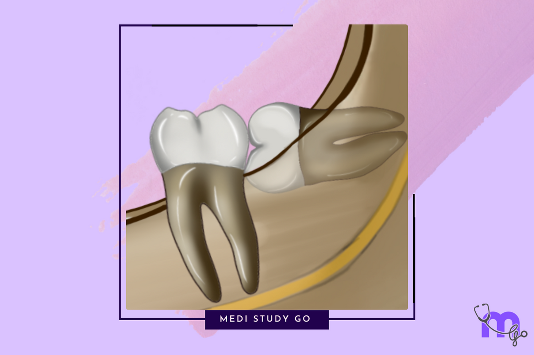
Related Resources
- Classification of Dental Impactions: A Detailed Guide for Dental Students
- Radiographic Assessment of Dental Impactions: Techniques and Interpretation
- Management of Impacted Teeth: Surgical Approaches and Techniques
- Complications of Dental Impactions: Prevention and Management
- Preventive Approaches to Dental Impactions: Early Intervention and Management
What is Dental Impaction?
Dental impaction refers to a condition where a tooth is prevented from erupting into its normal functional position within the dental arch. According to the World Health Organization (WHO), an impacted tooth is one that cannot erupt into its proper position due to obstruction by soft tissues, bone, or another tooth.
Impacted teeth are a common clinical challenge in dental practice, with third molar impaction being the most prevalent. If you're studying for your NEET exam, understanding the fundamentals of dental impactions is crucial for both theoretical knowledge and clinical application.
Why Do Teeth Become Impacted?
Several factors contribute to dental impactions:
- Lack of adequate space in the dental arch
- Abnormal eruption path or angulation
- Physical barriers like dense bone or adjacent teeth
- Genetic factors influencing tooth development
- Premature loss of primary teeth affecting eruption sequence
Understanding these underlying causes is essential for proper diagnosis and treatment planning, which is frequently tested in NEET previous year question papers.
Classification of Dental Impactions
Dental impactions are typically classified based on location and angulation. Let's explore the most common classifications you'll need to know for your NEET preparation.
Mandibular Third Molar Impactions
Mandibular third molars (wisdom teeth) are the most commonly impacted teeth. These impactions are classified using several systems:
Pell & Gregory Classification
This classification system considers two key factors:
-
Relationship to the Ramus of the Mandible:
- Class I: Sufficient space between the ramus and the second molar
- Class II: Reduced space where the crown is partially covered by the ramus
- Class III: No space available, with the crown fully enclosed in the ramus
-
Depth Relative to Occlusal Plane:
- Position A: Occlusal surface of the impacted tooth at the same level as the adjacent second molar
- Position B: Occlusal surface between the occlusal plane and cervical line of the second molar
- Position C: Occlusal surface below the cervical line of the second molar
Winter's Classification
This system classifies impactions based on the angulation of the third molar's long axis relative to the second molar:
- Mesioangular: Tilted toward the second molar (most common)
- Horizontal: Perpendicular to the second molar
- Vertical: Parallel to the second molar
- Distoangular: Tilted away from the second molar
- Inverted: Upside down with crown pointing toward the mandibular border
- Buccoversion: Tilted toward the cheek
- Linguoversion: Tilted toward the tongue
Maxillary Third Molar Impactions
Maxillary third molars also frequently become impacted. They are classified as:
- Vertical Impaction: Most common (63%)
- Distoangular Impaction: Second most common (25%)
- Mesioangular Impaction: Less common (12%)
- Horizontal Impaction: Rare
- Inverted Impaction: Very rare (1%)
Additionally, maxillary third molars may be categorized based on their proximity to the maxillary sinus:
- Sinus Approximation
- Non-Sinus Approximation
Maxillary Canine Impactions
Maxillary canines are the second most commonly impacted teeth. Their classification is based on position:
- Buccal Impaction: Canine positioned on the buccal surface
- Palatal Impaction: Canine positioned in the palate (more common)
- Alveolar Impaction: Canine positioned within the alveolar process
- Combined Impaction: Canine positioned in both palatal and labial aspects
- Impaction in Edentulous Maxilla
Radiographic Assessment of Impaction
Proper radiographic evaluation is crucial for accurate diagnosis and treatment planning of impacted teeth. Various imaging techniques assist in determining:
- Exact position and angulation of the impacted tooth
- Relationship to adjacent structures
- Presence of pathological conditions
- Anatomical variations
Radiographic Techniques
- Intraoral Periapical Radiographs (IOPA): Provides detailed view of the impacted tooth and surrounding structures
- Panoramic Radiographs: Offers a comprehensive view of the entire dentition
- Occlusal Radiographs: Helps in determining buccolingual position
- Cone Beam Computed Tomography (CBCT): Provides 3D visualization for complex cases
WHARFE Assessment
The WHARFE assessment by MacGregor evaluates the difficulty of extraction based on:
- W: Winter's classification (angulation)
- H: Height of the mandible
- A: Angulation of the third molar
- R: Root shape and development
- F: Follicle development
- E: Exit path availability
Predicting Inferior Alveolar Nerve Injury
The Rood and Shehab classification identifies radiographic signs that predict potential injury to the inferior alveolar nerve during extraction:
Root-Related Signs:
- Darkening of the root
- Deflected root
- Narrowing of the root
- Dark and bifid root apex
Canal-Related Signs:
- Interruption of white line
- Diversion of canal
- Narrowing of canal
Management Approaches
The management of impacted teeth depends on several factors including the type of impaction, patient's age, medical history, and presence of pathological conditions.
Indications for Removal of Impacted Mandibular Third Molars
- Recurrent pericoronitis
- Caries in the third or adjacent second molar
- Periodontal disease
- Pathological conditions (cysts, tumors)
- Unexplained pain
- Orthodontic considerations
- Pre-prosthetic considerations
Contraindications for Removal
- Advanced age (>35 years)
- Compromised medical status
- Proximity to vital structures with high risk of damage
- Asymptomatic deeply impacted teeth in older patients
- Potential for spontaneous eruption (in younger patients)
Surgical Procedure for Mandibular Third Molar Extraction
The typical procedure involves eight key steps:
- Aseptic Environment Preparation
- Regional Anesthetic Administration
- Flap Raising: Various designs including Ward's, modified Ward's, envelope, or triangular flaps
- Bone Removal: Using chisel and mallet, burs, or the "postage stamp" method
- Tooth Division: Based on the type of impaction
- Tooth Removal
- Wound Management
- Primary Closure
Lingual Split Bone Technique
This specialized technique is particularly useful for deeply impacted third molars and involves:
- Vertical Stop Cuts: Created at the mesial and distal aspects
- Buccal Cortical Plate Removal
- Distolingual Bone Fracturing
- Remaining Bone Removal
- Tooth Elevation
- Saucerization and Delivery
Complications of Impaction
Despite careful planning and execution, complications may occur during or after the extraction of impacted teeth. These include:
Intraoperative Complications
- Damage to adjacent teeth
- Fracture of the mandible
- Displacement of tooth fragments
- Injury to nerves (inferior alveolar, lingual)
- Excessive bleeding
- Soft tissue injury
Postoperative Complications
- Alveolar osteitis (dry socket)
- Infection
- Trismus (limited mouth opening)
- Paresthesia or anesthesia
- Postoperative pain and swelling
- Oro-antral communication (in maxillary impactions)
Understanding these complications and their management is essential for clinicians and students preparing for their NEET mock tests.
Preventive Measures
Several preventive approaches can reduce the incidence and complications of dental impactions:
- Early Orthodontic Intervention: Space maintenance and guidance of eruption
- Regular Dental Check-ups: Early detection of potential impactions
- Timely Extraction of Primary Teeth: When indicated
- Prophylactic Removal: In specific cases before complete root formation
- Patient Education: Importance of oral hygiene and regular follow-ups
These preventive strategies form an important part of comprehensive dental care and are crucial topics for NEET exam preparation.
Conclusion
Understanding dental impactions is essential for dental practitioners and students preparing for their NEET MDS. From classification systems to management techniques, this knowledge enables effective treatment planning and execution.
For more detailed information on specific aspects of dental impactions, explore our related articles below. If you're preparing for your NEET examination, our comprehensive guides provide valuable NEET tips and last-minute revision material to help you succeed.