
Oral Manifestations of Hemophilia: Clinical Recognition and Significance
Medi Study Go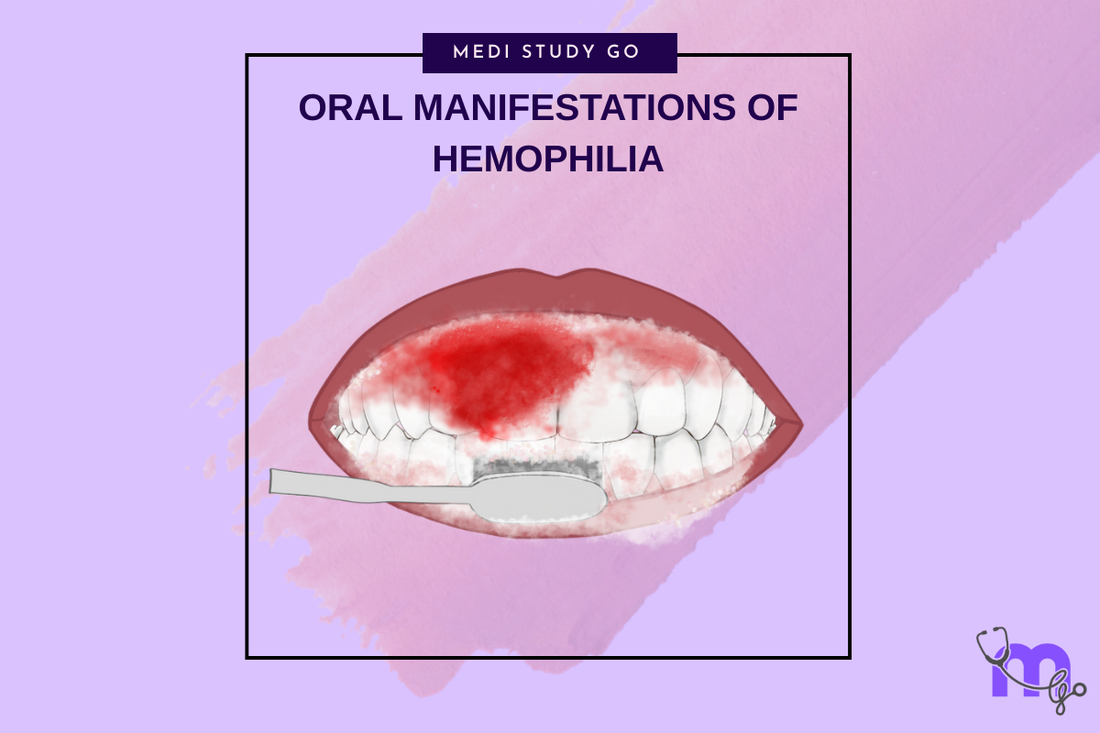
Related resources:
- Hemophilia Overview
- Dental Management Protocols
- Hemophilia Treatment Approaches
- Managing Complications
- Preventive Strategies
Understanding the oral manifestations of hemophilia is crucial for dental professionals to recognize potential undiagnosed cases and provide appropriate care for known patients with this bleeding disorder. This comprehensive guide examines the characteristic oral signs of hemophilia disease, their clinical significance, and implications for dental management.
Introduction to Hemophilia's Oral Presentation
Hemophilia manifests in the oral cavity in various forms, ranging from minor gingival bleeding to severe hemorrhagic episodes following dental procedures. Recognizing these signs is essential for dental practitioners, particularly those preparing for examinations like NEET MDS, as questions related to bleeding disorders frequently appear in NEET previous year question papers.
The oral cavity, with its rich vascular supply and frequent exposure to trauma during mastication and oral hygiene procedures, presents unique challenges in patients with clotting disorders. For individuals with hemophilia A and other coagulation deficiencies, even routine dental care can precipitate significant bleeding events without proper precautions.
Severe Gingival Bleeding: Hallmark Sign
Clinical Presentation and Recognition
One of the most common oral manifestations of hemophilia is disproportionate gingival bleeding following minimal trauma. This bleeding typically presents as:
- Spontaneous bleeding without apparent cause
- Prolonged bleeding after brushing or flossing
- Excessive bleeding during and after dental procedures
- Bleeding that is difficult to control with standard pressure techniques
Unlike typical gingivitis-related bleeding that resolves quickly with pressure, hemophilic bleeding may persist for hours or days without specialized intervention. The severity correlates with the patient's clotting factor levels, with severe hemophilia (factor levels <1%) presenting the highest risk for uncontrolled bleeding episodes.
Diagnostic Significance
Severe gingival bleeding disproportionate to the stimulus may serve as an early indicator of undiagnosed hemophilia, particularly in pediatric patients. Dental professionals should consider underlying bleeding disorders when encountering such presentations and obtain appropriate medical history and laboratory investigations. This knowledge is particularly valuable for students using NEET revision tools to prepare for clinical examinations.
Hemosiderin Deposits: Visual Indicator of Past Bleeding
Clinical Appearance
Repeated bleeding episodes in the oral tissues result in hemosiderin deposition, appearing as brownish-yellow discoloration of the gingiva and mucosa. These deposits represent the breakdown products of hemoglobin from extravasated blood and indicate a history of recurrent bleeding at the site.
In patients with hemophilia disease, hemosiderin deposits may be observed:
- Along the gingival margin
- In interdental papillae
- On the buccal mucosa adjacent to occlusal contact areas
- On dental hard tissues (causing brown discoloration of teeth)
Clinical Significance
The presence of hemosiderin deposits should alert dental practitioners to the possibility of a bleeding disorder even in the absence of active bleeding. These findings are particularly useful when evaluating patients with limited or unclear medical histories and should prompt further investigation into coagulation status.
Soft Tissue Manifestations: Petechiae, Ecchymosis, and Hematomas
Petechiae
Petechiae present as small (1-2mm), pinpoint hemorrhagic spots resulting from minor capillary bleeding. In patients with hemophilia, these commonly appear on:
- The soft palate
- Buccal mucosa
- Tongue
- Floor of the mouth
Unlike inflammatory lesions, petechiae do not blanch under pressure and may appear in clusters, particularly after minor trauma such as toothbrushing or consumption of rough-textured foods.
Ecchymosis
Larger than petechiae, ecchymoses (bruises) appear as diffuse areas of bluish-purple discoloration that may evolve to greenish-yellow as healing progresses. In the oral cavity, common sites include:
- The buccal mucosa (often along the occlusal line)
- Lateral borders of the tongue
- Lips (particularly following minor trauma)
- Floor of the mouth
The presence of ecchymoses without corresponding trauma history should raise suspicion for underlying hemophilia or other bleeding disorders.
Hematomas
Oral hematomas in hemophilia patients manifest as localized swellings containing extravasated blood. These can occur:
- In the tongue, potentially compromising airway
- In the buccal and labial mucosa following minor trauma
- In the floor of the mouth, which may lead to elevation of the tongue
- In the palate, particularly following dental injections or procedures
Hematomas require careful monitoring as they may continue to expand in patients with inadequate clotting factor levels, potentially leading to airway compromise in severe cases.
Pseudotumor of Hemophilia: Rare but Significant Finding
Clinical Presentation
The pseudotumor of hemophilia represents a rare but serious manifestation, appearing as a subperiosteal hematoma with reactive bone formation. In the maxillofacial region, these may present as:
- Slow-growing, painless swellings
- Radiographically visible bone changes
- Potential destruction of adjacent structures
- Resemblance to neoplastic lesions
Diagnostic Considerations
Pseudotumors require careful differential diagnosis from true neoplasms and other expansile lesions. Radiographic examination typically reveals a radiolucent area with reactive bone formation at the periphery. Biopsy is contraindicated due to the significant bleeding risk, making clinical correlation with hemophilia history and imaging findings essential for diagnosis.
Clinical Implications for Dental Management
Understanding these oral manifestations has direct implications for dental management strategies:
- Identification of unexplained oral bleeding should trigger investigation for potential hemophilia
- Presence of hemosiderin deposits warrants cautious approach to dental procedures
- Soft tissue manifestations indicate need for factor replacement before invasive procedures
- Pseudotumors require consultation with hematologist and oral surgeon
Importance for NEET Preparation
For dental students preparing for NEET MDS examinations, recognizing the oral manifestations of hemophilia represents an important clinical competency. This knowledge is frequently tested in various formats:
- Case-based questions in NEET q papers
- Clinical scenarios requiring differential diagnosis
- Treatment planning questions for patients with bleeding disorders
Using flashcard applications for NEET and NEET preparation books can help reinforce recognition of these characteristic presentations. Additionally, practicing with NEET mock tests builds confidence in identifying these manifestations in examination settings.
Conclusion
The oral manifestations of hemophilia provide valuable diagnostic clues for dental professionals. By recognizing severe gingival bleeding, hemosiderin deposits, petechiae, ecchymosis, hematomas, and pseudotumors, clinicians can identify patients requiring specialized management protocols and appropriate consultation with hematologists.
Early recognition of these signs facilitates safe delivery of dental care while preventing potentially life-threatening bleeding complications. For dental students and practitioners, this knowledge forms an essential component of comprehensive patient care and should be incorporated into clinical examination routines, particularly when evaluating patients with unclear bleeding histories or disproportionate bleeding responses.