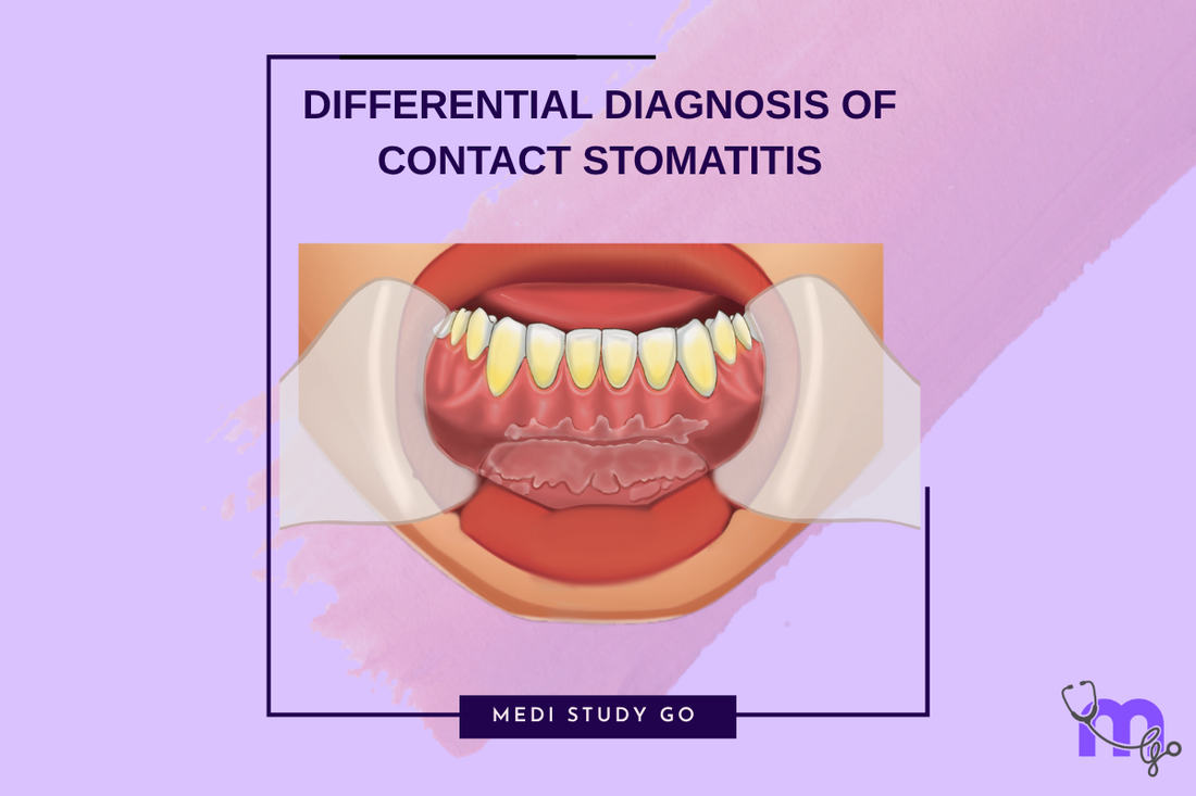
Differential Diagnosis of Contact Stomatitis
Medi Study Go
Quick Navigation to Specialized Topics:
- Complete Contact Stomatitis Guide
- What is Contact Stomatitis: Complete Definition, Causes
- Clinical Features & Types Detailed Analysis
- Treatment Protocols & Management
- How Long Does Contact Stomatitis Last
Introduction: Mastering the Art of Differential Diagnosis
Differential diagnosis questions represent approximately 30% of contact stomatitis-related items in NEET previous year question paper analysis, making this skill absolutely crucial for NEET MDS success. The ability to systematically distinguish allergic contact stomatitis from similar-appearing conditions often determines examination performance and clinical competence.
Quick Navigation to Specialized Topics:
- Understand Basic Mechanisms & Pathophysiology
- Master Clinical Feature Recognition
- Learn Evidence-Based Treatment Protocols
- Understand Recovery & Prognosis Patterns
- Complete Contact Stomatitis Guide Hub
This comprehensive differential diagnosis guide serves as your essential revision tool for NEET when tackling complex clinical scenarios. Whether you're analyzing NEET pyq patterns or preparing for viva voce examinations, this systematic approach ensures diagnostic accuracy and examination success.
Systematic Approach to Differential Diagnosis
Primary Decision Framework
Initial Assessment Questions:
- Location specificity: Does lesion correspond to contact area?
- Temporal relationship: Clear association with exposure/removal?
- Migration pattern: Does lesion remain fixed or move?
- Symmetry: Unilateral or bilateral involvement?
- Associated symptoms: Burning vs pain vs itching?
Risk Stratification:
- High probability contact stomatitis: Clear temporal + spatial relationship
- Moderate probability: Partial criteria met, requires further evaluation
- Low probability: No clear causative relationship, consider alternatives
Primary Differential Diagnoses

Contact Stomatitis vs Lichen Planus
This comparison frequently appears in NEET mock test scenarios and represents a high-yield topic for last minute revision.
Lichen Planus Distinguishing Features:
- Bilateral, symmetrical involvement (key differentiator)
- Wickham's striae in reticular pattern
- Multiple oral sites affected simultaneously
- Skin involvement often present
- No clear causative agent identified
Contact Stomatitis Distinguishing Features:
- Unilateral, asymmetrical pattern typically
- Corresponds exactly to contact area
- Clear causative agent identifiable
- Sharp, well-defined borders
- Resolution with allergen removal
Clinical Correlation Table:
| Feature | Lichen Planus | Contact Stomatitis |
|---|---|---|
| Distribution | Bilateral, symmetrical | Unilateral, contact-specific |
| Borders | Ill-defined, feathery | Sharp, well-demarcated |
| Wickham's striae | Classic reticular pattern | Peripheral striae only |
| Causative agent | Unknown/autoimmune | Identifiable allergen |
| Response to treatment | Slow, partial | Rapid with allergen removal |
Contact Stomatitis vs Leukoplakia
Leukoplakia Distinguishing Features:
- Cannot be wiped off (diagnostic criterion)
- No identifiable causative agent (idiopathic)
- Irregular, fuzzy borders common
- Possible malignant potential
- Predominantly in high-risk sites (lateral tongue, floor of mouth)
Contact Stomatitis Distinguishing Features:
- Clear relationship to causative agent
- Sharp, geometric borders especially with appliances
- Reversible condition with trigger removal
- Benign nature (no malignant potential)
- Location corresponds to contact pattern
High-Yield NEET Exam Tips:
- Leukoplakia = "Cannot wipe off" + "No clear cause"
- Contact stomatitis = "Sharp borders" + "Clear cause"
Contact Stomatitis vs Candidiasis
Pseudomembranous Candidiasis Features:
- White patches can be wiped off (pathognomonic)
- Underlying erythematous base revealed after wiping
- Predisposing factors (antibiotics, immunosuppression)
- Multiple oral sites typically affected
- Positive KOH test for fungal elements
Chronic Atrophic Candidiasis Features:
- Denture-bearing areas predominantly affected
- Ill-fitting dentures as predisposing factor
- Red, flat lesions without white component
- Burning sensation similar to contact stomatitis
- Antifungal response diagnostic
Key Differentiating Points:
- Contact stomatitis: Sharp borders, specific contact relationship
- Candidiasis: Predisposing factors, positive fungal testing
Advanced Differential Diagnoses
Contact Stomatitis vs Chemical Burns
Chemical Burns Characteristics:
- Immediate onset following exposure
- Severe tissue destruction often present
- Coagulation necrosis visible clinically
- Systemic symptoms possible
- Emergency treatment required
Contact Stomatitis Characteristics:
- Delayed onset (24-72 hours)
- Less severe tissue damage
- Inflammatory response without necrosis
- Localized symptoms only
- Gradual onset pattern
Clinical Correlation:
- Chemical burns: Immediate + Severe damage
- Contact stomatitis: Delayed + Inflammatory response
Contact Stomatitis vs Mucous Membrane Pemphigoid
Mucous Membrane Pemphigoid Features:
- Vesicles and bullae formation (rare in contact stomatitis)
- Positive Nikolsky's sign
- Gingival involvement predominant
- Ocular involvement possible
- Autoimmune etiology
Distinguishing Clinical Points:
- Pemphigoid: Bullae formation + Positive Nikolsky's
- Contact stomatitis: No bullae + Clear causative agent
Contact Stomatitis vs Erythema Multiforme
Erythema Multiforme Features:
- Target lesions on skin (pathognomonic)
- Lip involvement with crusting
- Acute onset with systemic symptoms
- Precipitating factors (drugs, infections)
- Self-limiting course
Key Differentiators:
- Erythema multiforme: Target skin lesions + Lip crusting
- Contact stomatitis: No skin involvement + Contact relationship
Location-Specific Differential Considerations
Gingival Lesions
When Contact Stomatitis Cinnamon Affects Gingiva:
- Horizontal band pattern along gingival margin
- Sharp demarcation at contact limits
- Unilateral involvement typically
Differential Considerations:
- Plaque-induced gingivitis: Poor oral hygiene, bilateral
- Necrotizing gingivitis: Painful, fetid odor, systemic symptoms
- Plasma cell gingivitis: Cobblestone appearance, bilateral
Buccal Mucosa Lesions
Cinnamon-Related Contact Stomatitis Patterns:
- Along occlusal plane distribution
- White, hyperkeratotic appearance
- Well-demarcated borders
Differential Considerations:
- Frictional keratosis: Irregular borders, chronic trauma history
- White sponge nevus: Bilateral, familial history, childhood onset
- Leukoedema: Disappears with stretching, bilateral
Restoration-Adjacent Lesions
Amalgam-Related Stomatitis:
- Does NOT migrate (pathognomonic feature)
- Sharp borders corresponding to restoration
- Chronic presentation typical
Differential Considerations:
- Oral squamous cell carcinoma: Irregular borders, indurated
- Traumatic ulcer: History of acute trauma, irregular shape
- Aphthous ulcer: Migrates, recurrent pattern, family history
NEET Previous Year Question Paper Analysis
High-Yield Question Patterns
Common Examination Scenarios:
Scenario 1: "A 45-year-old patient presents with bilateral red, edematous gingiva after changing toothpaste. Most likely diagnosis?"
Answer Approach:
- Identify bilateral involvement = systemic allergen
- Temporal relationship with toothpaste change
- Exclude: Lichen planus (no striae), gingivitis (poor hygiene absent)
- Confirm: Contact stomatitis due to dentifrice
Scenario 2: "White patches adjacent to amalgam restoration, present for 6 months, does not migrate. Diagnosis?"
Answer Approach:
- Non-migrating lesion = pathognomonic for amalgam stomatitis
- Chronic presentation (6 months)
- Exclude: Leukoplakia (would migrate), candidasis (different location)
- Confirm: Amalgam-related contact stomatitis
NEET Exam Tips for Differential Diagnosis
Memory Aids for Quick Recognition:
- "SHARP" borders = Contact stomatitis likely
- "BILATERAL" = Systemic allergen (dentifrice/mouthwash)
- "NON-MIGRATING" = Amalgam stomatitis
- "WIPEABLE" = Candidiasis (rules out contact stomatitis)
Systematic Elimination Strategy:
- Look for causative agent relationship first
- Assess border characteristics (sharp vs irregular)
- Check distribution pattern (unilateral vs bilateral)
- Consider temporal relationship (acute vs chronic)
- Evaluate response to treatment (rapid vs slow)
Advanced Diagnostic Considerations
When Multiple Conditions Coexist
Contact Stomatitis + Candidiasis:
- Immunocompromised patients at risk
- Overlapping symptoms (burning sensation)
- Sequential testing required (remove allergen first, then antifungal)
Contact Stomatitis + Lichen Planus:
- Rare but possible in susceptible individuals
- Different distribution patterns help distinguish
- Biopsy may be required for definitive diagnosis
Challenging Diagnostic Scenarios
Atypical Presentations:
- Multiple allergen exposure (complex patterns)
- Chronic, low-level exposure (subtle presentations)
- Concurrent conditions (overlapping features)
- Pediatric presentations (cooperation limitations)
When to Consider Biopsy:
- Uncertain diagnosis after clinical evaluation
- Atypical presentation patterns
- No response to appropriate treatment
- Malignancy concerns in high-risk patients
Flashcard Application for NEET Strategy
Effective Card Design
Front Side Questions:
- "Bilateral white striae, reticular pattern, no causative agent"
- "White patches, cannot wipe off, no clear cause"
- "Red gingiva, bilateral, new toothpaste history"
- "White lesion, adjacent to amalgam, does not migrate"
Back Side Answers:
- Lichen planus
- Leukoplakia
- Contact stomatitis (dentifrice)
- Contact stomatitis (amalgam)
Revision Tool for NEET Integration
Study Sequence:
- Learn individual features of each condition
- Practice comparison charts side-by-side
- Analyze clinical photographs systematically
- Test knowledge with flashcards
- Apply to NEET pyq scenarios
Conclusion: Diagnostic Excellence for NEET Success
Mastering differential diagnosis of allergic contact stomatitis requires systematic pattern recognition combined with understanding of key distinguishing features. The ability to rapidly eliminate incorrect options and identify pathognomonic features will ensure success in both NEET q paper examinations and clinical practice.